The frog - an introduction to anatomy and histology (1885) (14578875868)
_(14578875868).jpg?uselang=de)
Identifier: frogintroduction00mars (find matches)
Title: The frog : an introduction to anatomy and histology
Year: 1885 (1880s)
Authors: Marshall, A. Milnes, (Arthur Milnes. 1852-1893
Subjects: Frogs Amphibians Histology
Publisher: London : Smith, Elder
Contributing Library: Smithsonian Libraries
Digitizing Sponsor: Smithsonian Libraries
View Book Page: Book Viewer
About This Book: Catalog Entry
View All Images: All Images From Book
Click here to view book online to see this illustration in context in a browseable online version of this book.
Text Appearing Before Image:
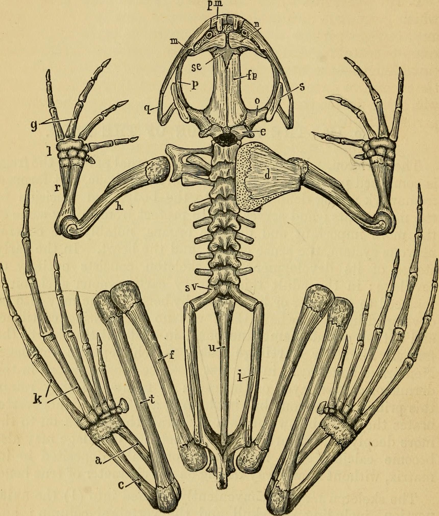
the first instance as ossifications in thedermis or deeper layer of the skin: in many fish they retainthis primitive position, but in the frog and most higher verte-brates they sink below the skin and graft themselves on to themore deeply placed cartilaginous skeleton. Cartilage may alsobecome calcified, i.e., have calcareous salts deposited in itsmatrix, without in any way taking on the character of true bone. The skeleton may be conveniently divided into (1) the axialportion, including the skull and the vertebral column : and(2) the appendicular portion, including the limbs and thelimb-girdles which attach them to the body. Examine the prepared skeletons and malce careful drawings toscale of the several parts. Colour, in your draivings, the cartilageblue, the cartilage hones yellow, and the membrane hones white orred. Prepare skeletons for yourself by soaking the parts in hotwater, and carefully brushing away the soft tissues until theskeleton is clean. 46 toE SitELEtON OP THE FROG.
Text Appearing After Image:
Fig. 7. The skeleton of the frog, seen from the dorsal surface: the leftsuprascapula and scapula have been removed. a, astragalus : c, calcaneum : d, suprascapula: e, exoccipital: /, femur :fip, frontoparietal:_ g, metacarpals : h, humerus : i, ilium : U, metatarsals :J, carpus : m, maxilla : n, nasal: o, pro-otic : %>, pterygoid : pm, premaxilla :g, quadratojugal: r, radio-ulna : s, squamosal: se, sphenethmoid: s.v,sacral vertebra : t, tibio-fibula : u, urostyle. THE VERTEBRAL COLUMN. 4^ A. The Axial Skeleton, I. The vertebral column or back bone. A bony tubewhich surrounds and protects the spinal cord: divisible intoan anterior part which is divided transversely into ninerings or vertebrae, and a posterior unsegmented portion of aboutequal length—the urostyle. At the sides of the tube betweenthe successive vertebra are the intervertebral foramina throughwhich the nerves pass out from the spinal cord to the variousparts of the body. a. Structure of a vertebra. Examine one of the
Note About Images


Relevante Bilder
.jpg)


Relevante Artikel
ExtremitätenevolutionAls Evolution der Extremitäten wird die Umwandlung der paarigen Flossen der Knochenfische (Osteichthyes) zu den Vorder- und Hintergliedmaßen der Landwirbeltiere (Tetrapoda) im Verlauf der Stammesgeschichte bezeichnet sowie der anschließende evolutionäre Formenwandel der Gliedmaßen nach dem Landgang der Wirbeltiere. .. weiterlesen