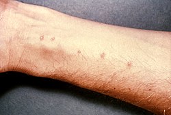
Schistosoma bladder histopathology
- Photo Credit:
- Content Providers(s): CDC/ Dr. Edwin P. Ewing, Jr.
Histopathology of bladder shows eggs of Schistosoma
by intense infiltrates of eosinophils and other inflammatory cells. Parasite.
|
Dieses Medium stammt aus der Public Health Image Library (PHIL), mit der Identifikationsnummer #34 der Centers for Disease Control and Prevention. Hinweis: Nicht alle PHIL-Bilder sind gemeinfrei; überprüfe unbedingt den Urheberrechtsstatus und die Nennung der Autoren und Inhaltsanbieter.
|
Relevante Bilder
Relevante Artikel
SchistosomiasisSchistosomiasis, auch Bilharziose, ist eine durch die Larven von Saugwürmern der Gattung Pärchenegel (Schistosoma) verursachte Wurmerkrankung. Sie wird in warmen Binnengewässern durch Schnecken als Zwischenwirte verbreitet. .. weiterlesen


