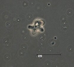
SEM image of ultrastructural morphology exhibited by a number of Hartmannella vermiformis amoebae trophozoites
Autor/Urheber:
Janice Haney Carr
Shortlink:
Quelle:
Größe:
700 x 475 Pixel (84876 Bytes)
Beschreibung:
Magnified 1155X, this 2002 scanning electron microscopic (SEM) image revealed some of the ultrastructural morphology exhibited by a number of Hartmannella vermiformis amoebae trophozoites. As part of a study to determine whether Legionella pneumophila bacteria can colonize, and grow in biofilms with, or without the presence of H. vermiformis, here these protozoa were situated atop a base biofilm composed of Pseudomonas aeruginosa, Klebsiella pneumoniae and a Flavobacterium sp. bacteria. Note that the amoebae were grazing upon these bacteria, the end result of which can be seen in PHIL 11166, which reveals the scoured stainless steel coupon surface. See the link below for more on this study.
Note: This species has been re-classified as Vermamoeba vermiformis by Smirnov et al., 2011. doi:10.1016/j.protis.2011.04.004, doi:10.3389/fmicb.2022.808499.
Note: This species has been re-classified as Vermamoeba vermiformis by Smirnov et al., 2011. doi:10.1016/j.protis.2011.04.004, doi:10.3389/fmicb.2022.808499.
Lizenz:
Public domain
Credit:

|
Dieses Medium stammt aus der Public Health Image Library (PHIL), mit der Identifikationsnummer #11166 der Centers for Disease Control and Prevention. Hinweis: Nicht alle PHIL-Bilder sind gemeinfrei; überprüfe unbedingt den Urheberrechtsstatus und die Nennung der Autoren und Inhaltsanbieter.
|
Relevante Bilder
Relevante Artikel
Vermamoeba vermiformisVermamoeba vermiformis ist eine Art (Spezies) von Amöben (Amoebozoa) in der Gruppe Echinamoebida der Tubulinea. .. weiterlesen


