Picomonas judraskeda
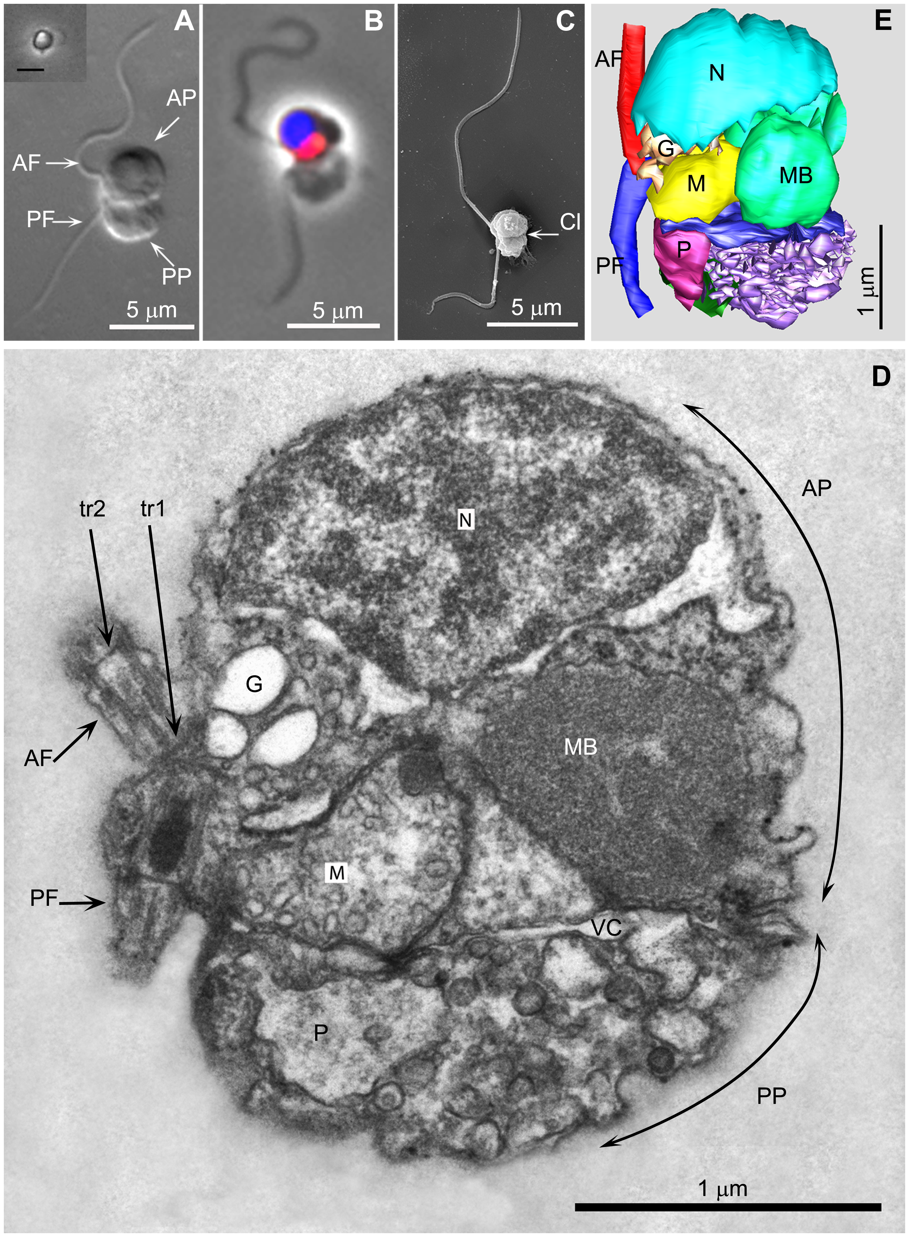
- A. Differential interference contrast of a chemically fixed cell. Inset shows phase contrast image of a live cell from tissue culture flask photographed with an inverted microscope (Scale bar 5 µm).
- B. Fluorescence and phase contrast overlay, nucleus (blue), mitochondrion (red).
- C. SEM image.
- D. A longitudinal section through a cell in the plane of the flagella, viewed from the cell’s left.
- E. A 3 D serial section reconstruction of the cell depicted in 2D. AF/PF (anterior−/posterior flagellum); AP/PP (anterior/posterior part of the cell); G (Golgi body); M (mitochondrion); MB (‘microbody’); N (nucleus); tr1,tr2 (distal [tr2] and proximal [tr1] flagellar transitional regions); P (posterior digestive body); Cl (cleft separating the anterior from the posterior part of the cell); vc (vacuolar cisterna).
An den Hochlader:: Bitte unbedingt einen Link (URL) zur Original-Datei oder zum dazugehörigen Artikel angeben.


Relevante Bilder
_(1910)_(17950796051)-9%2b10%2b11.jpg)


.png)


Relevante Artikel
PicozoaDie Picozoa sind ein im Jahr 2013 neu beschriebener Stamm heterotropher Einzeller aus der Domäne der Eukaryoten. Sie sind die kleinsten Formen frei im Wasser schwebender Lebewesen, werden zwischen einem halben und 3,8 µm (Mikrometer) groß und kommen in großer Zahl und Dichte in allen Weltmeeren vor. .. weiterlesen
HacrobiaDie Kryptomonaden-Haptophyten-Gruppe ist eine vorgeschlagene monophyletische Gruppierung (Klade) von einzelligen Eukaryoten, die nicht zur SAR-Supergruppe gehören. Mehrere alternative Namen wurden für diese Gruppe verwendet, darunterHacrobia, CCTH ; sowie Eukaryomonadae. .. weiterlesen