Guide leaflet (1901) (14763631314)
_(14763631314).jpg?uselang=de)
Identifier: scienceguide1630amer (find matches)
Title: Guide leaflet
Year: 1901 (1900s)
Authors: American Museum of Natural History
Subjects: American Museum of Natural History Natural history
Publisher: New York : The Museum
Contributing Library: American Museum of Natural History Library
Digitizing Sponsor: IMLS / LSTA / METRO
View Book Page: Book Viewer
About This Book: Catalog Entry
View All Images: All Images From Book
Click here to view book online to see this illustration in context in a browseable online version of this book.
Text Appearing Before Image:
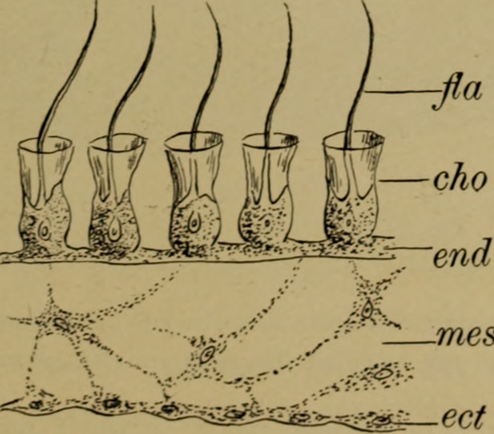
ionally the characteristics of amouth. The walls of the vase are perforated with numerous regularlyarranged openings or pores (p.) which open directly into thehollow interior of the sponge—called the paragastric or atrialcavity (atr.). The walls are made up of three layers: 1st, theectoderm, or outer layer; 2d, the endoderm, or inner layer;3d, the mesoderm, or middle layer. The ectoderm (ect.) is a thin layer of cells, generally arrangedin mosaic form and known as pavement cells. In the case ofthis species, however, the walls of the cells have disappearedand left the protoplasmic cell-contents continuous over theentire surface of the animal. Such a layer is called a syncytium.The endoderm (end.) lines the paragastric cavity and is madeup of a layer of peculiar and characteristic cells called collaredcells, or choanocytes (cho.),found nowhere else among many-celledanimals. They are so called from a collar-like rim around theouter edge of the cell out of which extends a long whip-like
Text Appearing After Image:
lues FIG 8.—SECTION THROU3H SPONGE WALLect., ectoderm ; mes.% mesoderm ; em/., endoderm \cho.y choanocytes or collared cells;Jla., flagellum. filament or flagellum (fla.). The continuous vibration of theseflagella produces a current by means of which the sea-water,with its multitude of tiny animal and plant forms, is sucked inthrough the pores. The organisms are then seized upon by the (9) 224 THE AMERICAN MUSEUM JOURNAL choanocytes and their digestible parts absorbed. What is leftis discarded and flows with the current out through the osculumat the summit of the vase. The mesoderm (mes.) is a thin jelly-like layer between theectoderm and endoderm. It contains scattered amoeboid cellsand the reproductive elements, and is the origin of the skeleton. 2. The Canal Systems. In the form of sponge just described the mesoderm isextremely thin, but if, as in the majority of sponges, there is agreater or less thickening of this layer, the pores will no longerbe perforations, but will become
Note About Images


Relevante Bilder

Relevante Artikel
ChoanocytChoanocyten sind ein Zelltyp, der bei Schwämmen (Porifera) vorkommt. Die Choanozyten bilden in den Schwämmen eine Deckzellschicht, die Choanoderm genannt wird. Das Choanoderm kleidet den Innenraum der Schwämme aus. Die Kragengeißelzelle besteht aus einem rundlichen Zellkörper (Soma). An einem Zellpol sitzt eine lange Geißel (Flagellum). Um die Geißel herum steht ein Ring aus Stereovilli. Dies ist der namensgebende Kragen. Zwischen den einzelnen Stereovilli sitzt Schleim. So entsteht ein Schleimsaum (Plasmakragen). Durch den Geißelschlag wird ein Wasserstrom erzeugt, der von den Seiten Wasser zuführt. Das Wasser tritt durch den Schleimsaum. Im Wasser schwebende Nahrungspartikel verfangen sich im Schleim. Sie werden anschließend durch Pseudopodien eingefangen und mittels Endozytose von der Kragengeißelzelle aufgenommen. .. weiterlesen