Fmicb-05-00003-g001D-p2
Autor/Urheber:
Silvia Spinelli, David Veesler, Cecilia Bebeacua, Christian Cambillau
Attribution:
Das Bild ist mit 'Attribution Required' markiert, aber es wurden keine Informationen über die Attribution bereitgestellt. Vermutlich wurde bei Verwendung des MediaWiki-Templates für die CC-BY Lizenzen der Parameter für die Attribution weggelassen. Autoren und Urheber finden für die korrekte Verwendung der Templates hier ein Beispiel.
Shortlink:
Quelle:
Größe:
284 x 700 Pixel (69897 Bytes)
Beschreibung:
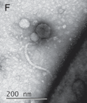
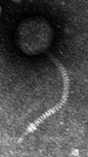
Lactococcal siphophage p2, generated by assembling the reconstructions of the capsid (top), connector and tail (middle), and the tail-tip (bottom) on low-resolution maps of the full phages. In the capsid map, pentons are identified by red arrows/points and hexons by green arrows/points. Dimensions are given in Å and the angle of rotation between MTP hexamers is given in degrees.
Lizenz:
Credit:
Structures and host-adhesion mechanisms of lactococcal siphophages. In: Front. Microbiol., Volume 5 (2014), Sec. Virology, Research Topic Gram-positive phages: From isolation to application. doi:10.3389/fmicb.2014.00003.
Relevante Bilder
Relevante Artikel
SiphovirenDie morphologisch begründete (nicht-taxonomische) Gruppe der Siphoviren umfasst eine Reihe von Familien, Unterfamilien und Gattungen von Viren mit einem linearen Molekül doppelsträngiger DNA (dsDNA) als Genom von ca. 22–121 kbp Länge. .. weiterlesen


