A system of gynecology (1887) (14596290390)
_(14596290390).jpg?uselang=de)
Identifier: system01mann (find matches)
Title: A system of gynecology
Year: 1887 (1880s)
Authors: Mann, Matthew D. (Matthew Darbyshire), 1845-1921
Subjects: Women
Publisher: Philadelphia, Lea brothers & co.
Contributing Library: Yale University, Cushing/Whitney Medical Library
Digitizing Sponsor: Open Knowledge Commons and Yale University, Cushing/Whitney Medical Library
View Book Page: Book Viewer
About This Book: Catalog Entry
View All Images: All Images From Book
Click here to view book online to see this illustration in context in a browseable online version of this book.
Text Appearing Before Image:
een described, blends anteriorly withthe muscles above mentioned ; some of its peripheral fibres are appa-rently continuous with those of the bulbo-cavernosi. The pubo-coccygeus (anterior portion of the levator ani), as viewed from below,lies deeper than the preceding muscles (/. c. above them), as it is behindthe perineal septum (Fig. 78). It encircles the vagina, and its innerfibres curve inward behind that canal to enter the perineal body behindthe lower edge of the septum. When traced farther backward they sur-round the rectum in a similar manner between the two sphincters, andblend with the terminal fibres of the longitudinal laver of the rectum. THE PERINEAL BODY. 231 The erectores clitoridis are not properly included in this dissection,and are described with the clitoris. Removing the bulbo-cavernosi and the vaginal bulbs, which restupon the anterior layer of the triangular ligament, the latter is seento he perforated by branches of the pudic arteries and nerves and by Fig. 78.
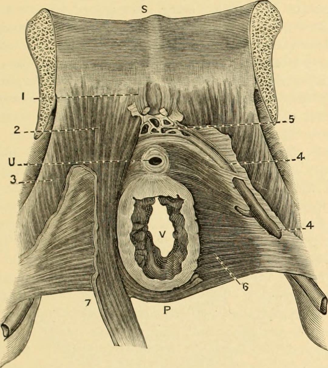
Text Appearing After Image:
Perineal Septum, posterior view (Savage); S, posterior surface of symphysis; U, urethra; Vtvagina; l. pubic attachment of bladder; 2, pubic attachment of levatorani (pubo-coccyg-eus); ■■.lino of attachment of obturato-coccygeus; 1, pudic vein; 5, urethro-pubic plexusof veins; 6, posterior surface of septum; 7, median portion of pubo-coccygeus, enteringperinea) body at lower edge of septum. the communicating veins which extend from the bulbs and clitoristo the vesical plexuses. When the layer itself is detached, the follow-in- structures are exposed : The urethra, surrounded by the compressorurethra1 of Guthrie, the constrictor vaginae of some authors;2 the deeptransversus perinei, the vulvo-vaginal glands, internal pudic vesselsand nerves, dorsal vein and nerve of the clitoris, and artery of thebulb. The two former muscles are described by Heath as forming afigure-of-8 around the urethra and vagina, being attached anteriorly tothe posterior aspect of the pubic arch, and entering t
Note About Images


Relevante Bilder


Relevante Artikel
BeckenbodenDer Beckenboden ist der bindegewebig-muskulöse Boden der Beckenhöhle beim Menschen. Er wird unter anderem durch den Musculus levator ani gebildet. Auf Grund der unterschiedlichen Körperhaltung und Beckenstellung bezeichnet der Begriff Beckenboden bei den vierfüßigen Säugetieren die von Scham- und Sitzbein gebildete Ventralfläche des knöchernen Beckens. Der dem Beckenboden des Menschen entsprechende hintere Abschluss der Beckenhöhle wird als retroperitonealer Teil der Beckenhöhle bezeichnet. .. weiterlesen