A computed tomography brain scan showing bilateral basal ganglia calcification
Autor/Urheber:
Abu-Amero KK, Al-Dhalaan H, Bohlega S, Hellani A, Taylor RW.
Attribution:
Das Bild ist mit 'Attribution Required' markiert, aber es wurden keine Informationen über die Attribution bereitgestellt. Vermutlich wurde bei Verwendung des MediaWiki-Templates für die CC-BY Lizenzen der Parameter für die Attribution weggelassen. Autoren und Urheber finden für die korrekte Verwendung der Templates hier ein Beispiel.
Shortlink:
Quelle:
Größe:
1200 x 883 Pixel (156919 Bytes)
Beschreibung:
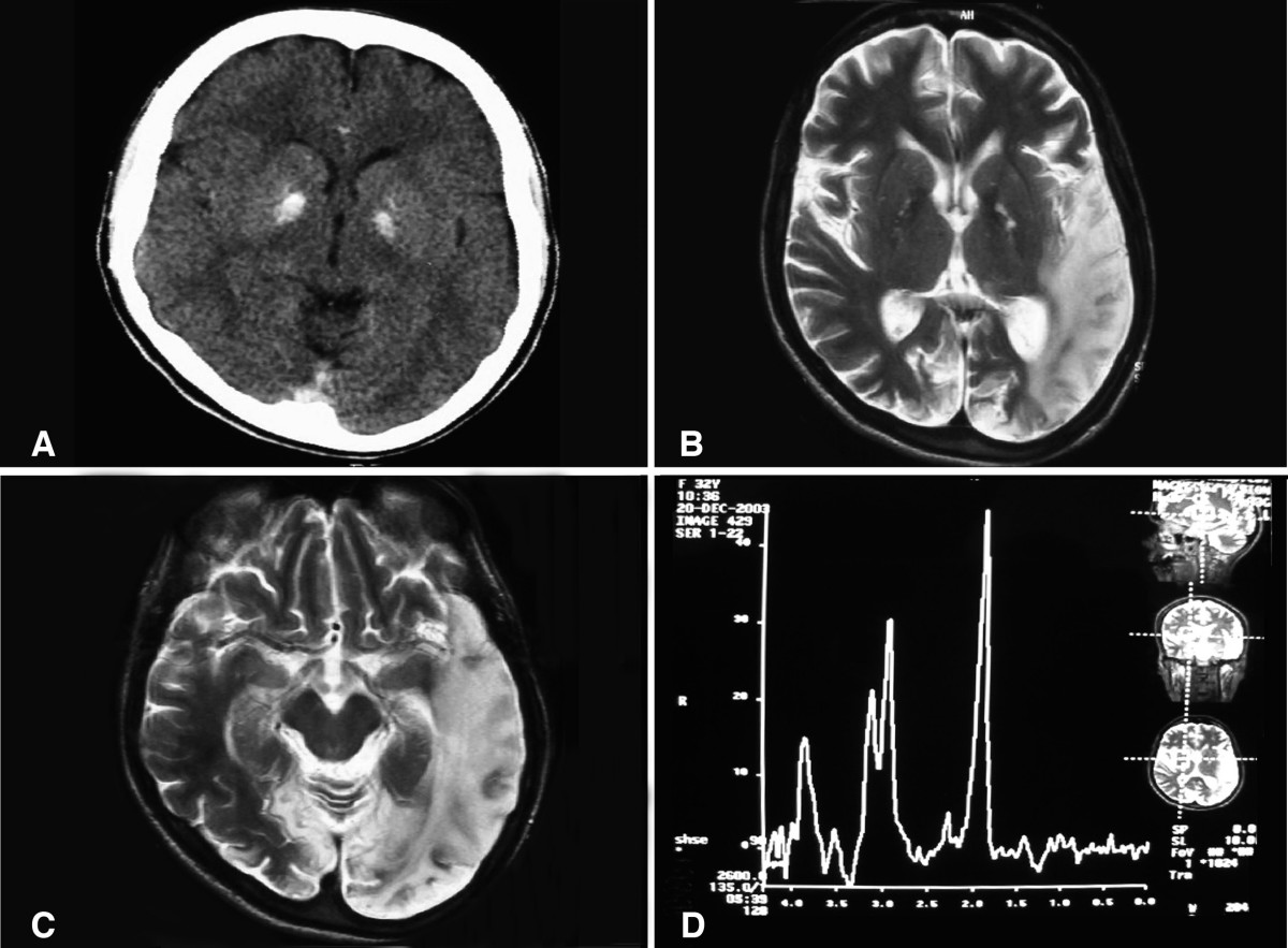
(a) A computed tomography brain scan showing bilateral basal ganglia calcification; the cerebellum shows prominent folia indicating mild cerebellar atrophy. (b) Axial T2 brain magnetic resonance image scan showing left temporo-parieto occipital ischemic lesion. (c) Axial T2 brain magnetic resonance image scan showing the extension of the parietal temporal region to the occipital lobe, and also showing a right occipital lesion. (d) Magnetic resonance spectroscopy showing inversion of J-coupling phenomenon at 1.3 ppm, indicating lactate peak. Abu-Amero et al. Journal of Medical Case Reports 2009 3:77 doi:10.1186/1752-1947-3-77
Kommentar zur Lizenz:
© 2009 Abu-Amero et al; licensee BioMed Central Ltd.
This is an Open Access article distributed under the terms of the Creative Commons Attribution License (https://creativecommons.org/licenses/by/2.0), which permits unrestricted use, distribution, and reproduction in any medium, provided the original work is properly cited.
Lizenz:
Bild teilen:


Relevante Bilder

Relevante Artikel
Aicardi-Goutières-SyndromDas Aicardi-Goutières Syndrom (AGS) ist eine seltene angeborene Erkrankung des Gehirns mit den Hauptmerkmalen einer subakuten Enzephalopathie mit Kalkablagerungen in den Basalganglien, Leukodystrophie und Lymphozytose der Zerebrospinalflüssigkeit (CSF). .. weiterlesen