6nb6 prefusion 6m3w postfusion spike
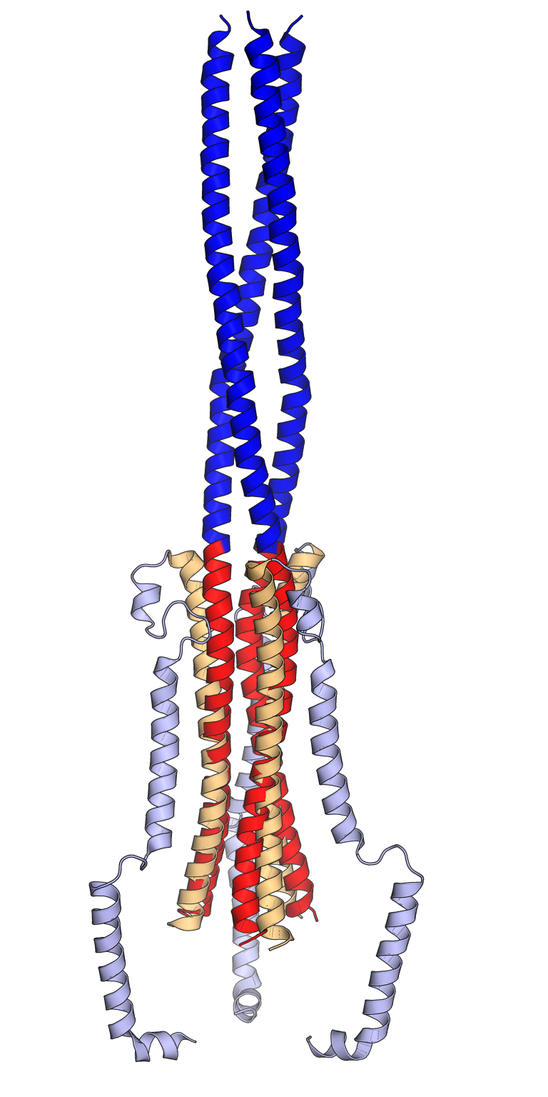
Comparison of the pre-fusion (orange, light blue) and post-fusion (red, dark blue) conformations of the SARS-CoV spike protein trimer. In the pre-fusion conformation, the central helix (orange) and heptad repeat 1 (HR1, light blue) are folded back on each other in an antiparallel orientation. In the post-fusion conformation, the central helix (red) and the HR1 sequence (dark blue) reorganize to form an extended trimeric coiled coil. The viral membrane is at the bottom and the host cell membrane at the top. Only key portions of the S2 subunit are shown. Rendered using PyMol from cryo-electron microscopy structures PDB: 6NB6 (pre-fusion) and PDB: 6M3W (post-fusion) superposed using the central helix sequences, inspired by Figs 1 and 2 from Fan 2020.
6NB6: Unexpected Receptor Functional Mimicry Elucidates Activation of Coronavirus Fusion. Walls, A.C., Xiong, X., Park, Y.J., Tortorici, M.A., Snijder, J., Quispe, J., Cameroni, E., Gopal, R., Dai, M., Lanzavecchia, A., Zambon, M., Rey, F.A., Corti, D., Veesler, D. (2019) Cell 176: 1026-1039.e15
PubMed: 30712865 DOI: 10.1016/j.cell.2018.12.028
6M3W: Cryo-EM analysis of the post-fusion structure of the SARS-CoV spike glycoprotein. Fan, X., Cao, D., Kong, L., Zhang, X. (2020) Nat Commun 11: 3618-3618
PubMed: 32681106 DOI: 10.1038/s41467-020-17371-6


Relevante Bilder


Relevante Artikel
Spike-Glykoprotein von SARS-CoV-2Das Spike-Glykoprotein von SARS-CoV-2 ist eine nach außen ragende Proteinstruktur (Peplomer) des SARS-CoV-2 Virions. Es ist ein transmembranes Glykoprotein auf der Virusoberfläche des SARS-CoV-2 und dient als Ligand zum Andocken an ACE2 auf der Zelloberfläche sowie als fusogenes Protein zum Zelleintritt. Wie bei den anderen Mitgliedern der Coronaviridae bilden die Spike-Gykoproteine die markante und namensgebende Oberflächenstruktur dieser Virionen. .. weiterlesen