Squirmida
| Squirmida | ||||||||||||
|---|---|---|---|---|---|---|---|---|---|---|---|---|
 DIC-Aufnahme lebender Trophozoiten von Platyproteum vivax | ||||||||||||
| Systematik | ||||||||||||
| ||||||||||||
| Wissenschaftlicher Name ohne Rang | ||||||||||||
| Squirmidea | ||||||||||||
| Cavalier-Smith, 2014[1] | ||||||||||||
| Wissenschaftlicher Name ohne Rang | ||||||||||||
| Squirmida | ||||||||||||
| Cavalier-Smith, 2014[1] |

Squirmida ist ein Taxon von Einzellern innerhalb der Alveolata. In Taxonomien der Protisten mit Rangstufen haben die Squirmida den Rang einer Ordnung und sind die einzige Ordnung in der Klasse Squirmidea. Zu den Squirmida werden drei Gattungen klassifiziert (Stand Januar 2025): Filipodium, Platyproteum und Digyalum, die früher der Gregarinen-Familie Selenidiidae innerhalb der parasitischen Apicomplexa zugeordnet waren.
Die Squirmida (und Squirmidea) wurden 2014 vom Protistologen Thomas Cavalier-Smith nach phylogenetischen Untersuchungen zunächst für die beiden Gattungen Filipodium und Platyproteum eingerichtet. Da frühere Maximum-Likelihood-Bäume zu keinen befriedigenden Ergebnissen geführt hatten, wurde jetzt mit einem neuen Modell, das ungleichmäßigsten Raten der rDNA-Evolution in standortheterogenen rDNA-Bäumen umfasste, 122 Gregarinen (nach damaliger Taxonomie) und 452 Außengruppen erfasst. Dabei zeigte sich, dass die damalige große Gruppe der Gregarinen polyphyletisch war, was neben anderen Verschiebungen zunächst zur Ausgliederung der beiden Gattungen in die neue Ordnung Squirmida führte.[1] Weitere Untersuchungen von Jan Janouškovec et al. zeigten 2019, dass auch die verbleibenden Apicomplexa noch polyphyletisch waren und führten u. a. zur Ausgliederung der Gattung Digyalum (Filipodium und Platyproteum wurden hier nicht erfasst).[3] Inzwischen (Stand Februar 2024) wurde diese Gattung ebenfalls den Squirmida zugeordnet.[2]
Systematik
Die Systematik der Squirmida ist daher wie folgt (Stand Februar 2024 bis Januar 2025):[2]
Klasse Squirmidea Cavalier-Smith, 2014[1]
- Ordnung Squirmida Cavalier-Smith, 2014[1]
- ohne Familenzuweisung
- Familie Filipodiidae Cavalier-Smith, 2014[1]
- Gattung Filipodium Hukui 1939[7][8]
- Familie Platyproteidae Cavalier-Smith, 2014[1][9]
- Gattung Platyproteum Rueckert & Leander 2009[9][10]
Filipodium
Filipodium ist eine Gattung von mikroeukaryotischer Parasiten, die 1939 von Tosito Hukui erstbeschrieben wurde.[7] Die Gattung wurde zunächst innerhalb der Gruppe der Gregarinen (Apicomplexa) in die Familie Lecudinidae gestellt.[7] Sie wurde später in die Gregarinen-Familie Selenidiidae überführt, als sich ihre verwandtschaftliche Nähe zu Platyproteum vivax herausstellte, denn sie bildeten eine englisch squirmids genannte Klade, die in Maximum-Likelihood-Bäumen schwach mit Selenidium terebellae gruppiert war (Rueckert und Leander 2009).[9][8] Diese Klade mit beiden Gattungen wurden dann 2014 von Cavalier-Smith in die neu eingerichtete Gruppe (Ordnung) Squirmida außerhalb der Apicomplexa ausgelagert.[1][2]
Die Typusart der Gattung ist Filipodium ozakai, ihr Wirt ist der Spritzwurm Siphonosom – alle Arten dieser Gattung befallen wirbellose Meerestiere.[7]
Beschreibung
Das Mucron[A. 1] ist breit und trichterförmig mit Papillen am Rand. Die Gamonten sind länglich, längsgestreift und haben viele abstehende Filamente, die unter der Pellicula hervortreten. Die Gametozysten (Zystenstadium der Gameten) haben zahlreiche Oozysten. Die Gameten sind unterschiedlich; aber wie die weiblichen haben auch die männlichen Gameten haben keine Geißel. Die Oozysten sind ellipsoidisch oder eiförmig und haben 8 Sporozoiten.[7][9][1]
Lebenszyklus
Die Typusart infiziert die Sipunculida (Spritzwürmer). Der Parasit infiziert den Magen-Darm-Trakt des Wirts und wird vermutlich über den orofäkalen Weg (durch mit der Mundöffnung des Wurms aufgenommene Fäkalien der Artgenossen) übertragen, aber die Einzelheiten dieses Mechanismus sind derzeit noch unbekannt.
Platyproteum


Platyproteum ist eine Gattung parasitischer Alveolata aus der Verwandtschaft der Apicomplexa (d. h. der Myzozoa). Die Gattung wurde 2009 von Sonja Rueckert und Brian Leander erstbeschrieben. Die Typusart aus der Erstbeschreibung ist Platyproteum vivax, ihr Wirt der Spritzwurm Phascolosoma agassizii. Diese Art war in ihrer Erstbeschreibung von Gunderson & Small 1986 der Gregarinen-Gattung Selenidium zugeordnet worden, aber dann 2009 durch Rueckert und Leander in die neue Gattung Platyproteum ausgegliedert.[9] Es besteht eine verwandtschaftliche Nähe zu Filipodium phascolosomae, die beiden Gattungen bilden eine zunächst englisch squirmids genannte Klade innerhalb der Gregarinen-Familie Selenidiidae,[9] die 2014 von Cavalier-Smith zur Ordnung Squirmida (außerhalb, aber in verwandtschaftlicher Nähe der Apicomplexa) erklärt wurde.[1]
Die Arten dieser Gattung sind bandförmige Parasiten, die wirbellose Meerestiere infizieren.[9]
Lebenszyklus
Der Parasit infiziert den Magen-Darm-Trakt des Wirts und wird vermutlich wie bei der Gattung Filipodium über den orofäkalen Weg übertragen, aber auch hier sind die Einzelheiten dieses Mechanismus derzeit noch unbekannt.[9]
Bildergalerie
- Platyproteum vivax
 DIC-Aufnahmen lebender Trophozoiten. Vorderende einer Zelle mit zwei kurzen Geißeln (Doppelpfeile). Balken: 10 µm.
![REM-Aufnahmen des „Vorderen Apparats“ (englisch anterior apparatus)[A. 1] mit vorderer und hinterer Geißel (Af respektive PF). Balken: 2 µm.](//upload.wikimedia.org/wikipedia/commons/thumb/7/7a/Botany-ubc-bleander-Platyproteum2024-Fig3DE.jpg/500px-Botany-ubc-bleander-Platyproteum2024-Fig3DE.jpg)

 Aufnahme eines Trophozoiten, rot zeigt α-Tubulin an. N: Zellkern, Balken: 20 µm.
 Konfokale LSM-Aufnahme von zweier verschieden gestreckter Trophozoiten, die vielen Mitochondrien grün gefärbt (unter der Zellmembran sitzend). Balken: 20 µm
Digyalum
Digyalum ist eine Gattung mariner Alveolata, die von Koura et al. Anfang der 1990er Jahre mit einer einzigen Spezies Digyalum oweni erstbeschrieben wurde und von diesen Autoren ursprünglich als „aseptate Gregarine“ der Familie Selenidiidae Brasil, 1907 zugeordnet wurde.[4][5][6] Digyalum oweni wurde aus dem Darm der Strandschnecken-Art Littorina obtusata isoliert. Von Anfang an war deutlich, dass sie eine für Gregarine ungewöhnliche Eigenschaft hat, indem dass die Falten auf der Zelloberfläche quer verlaufen und die Zellen zwei anteriore (vordere) beutelartige Vertiefungen aufweisen.[4]
Die Gattung wurde von Janouškovec et al. 2019 nach weiteren Analysen von den Gregarinen abgetrennt und außerhalb der Apicomplexa, jedoch in verwandtschaftlicher Nähe zu ihnen angesiedelt.[3] Inzwischen ist klar, dass sie zu der bereits bestehenden und ebenfalls in diesem Bereich befindlichen Ordnung Squirmida gehört.[2]
Beschreibung
Die einzige Spezies und Typusart Digyalum oweni ist ein den Parasit im Darm einiger Arten der marinen Strandschnecken-Gattung Littorina. Das vordere Anheftungsorgan (Mucron)[A. 1] besteht aus einem Ring von Lappen (englisch lobes) plus einem Ring aus körnigem (granularem) Material, der in die Spitze der Wirtsdarmzelle eingebettet wird. Die Wirtszelle wird zwar in gewissem Maße geschädigt, doch beschränkt sich dies auf die Anheftungsstelle, ansonsten scheint der Parasit der Schnecke nicht wesentlich zu schaden. Neben der Anheftung des Parasiten an seinem Wirt spielt das Mucron möglicherweise auch eine Rolle bei der Ernährung und als Mikrotubuli-Organisationszentrum.[14]
Anmerkungen
- ↑ a b c d Das Mucron ist ein „Anheftungsorganell“ (englisch attachment organelle) der Archigregarinen und phänotypisch ähnlicher epizellulärer (an der Außenseite der Wirtszellen) parasitischer Alveolata aus der Apicomplexa-Verwandtschaft (Myzozoa). Man nimmt an, dass es sich vom apikalen Komplex ableitet.[11][12][13] Offensichtlich bezeichnet der Ausdruck dasselbe, was bei anderen Autoren „Vorderer Apparat“ (englisch anterior apparatus) genannt wird – vielleicht, weil der Ausdruck „Mucron“ den eigentlichen Gregarinen vorbehalten bleiben soll.
Einzelnachweise
- ↑ a b c d e f g h i j Thomas Cavalier-Smith: Gregarine site-heterogeneous 18S rDNA trees, revision of gregarine higher classification, and the evolutionary diversification of Sporozoa. In: European Journal of Protistology. 50. Jahrgang, Nr. 5, Oktober 2014, S. 472–495, doi:10.1016/j.ejop.2014.07.002, PMID 25238406 (englisch).
- ↑ a b c d e f g Danja Currie-Olsen, Brian S. Leander: Novel cytoskeletal traits in the intestinal parasites (Squirmida, Platyproteum vivax) of Pacific peanut worms (Sipuncula, Phascolosoma agassizii). In: Journal of Eukaryotic Microbiology, Band 71, Nr. 3, 25. Februar 2024, S. e13023; doi:10.1111/jeu.13023 (englisch) Siehe insbes. Fig. 1.
- ↑ a b c Jan Janouškovec, Gita G. Paskerova, Tatiana S. Miroliubova, Kirill V. Mikhailov, Thomas Birley, Vladimir V. Aleoshin, Timur G. Simdyanov: Apicomplexan-like parasites are polyphyletic and widely but selectively dependent on cryptic plastid organelles. In: eLife, Band 8, 16. August 2019; doi:10.7554/ELIFE.49662, ISSN 2050-084X, PMC 6733595 (freier Volltext), PMID 31418692 (englisch). Siehe insbes. Fig. 1.
- ↑ a b c d E. A. S. Koura, John W. Grahame, R. W. Owen, Esra G. Kamel: Digyalum oweni, gen. nov., sp. nov., a new and unusual gregarin protozoan from the gut of mollusc Littorina obtusata (Prosobranchia: Gastropoda). In: Journal of the Egyptian Society of Parasitology, Band 20, Nr. 1, Juni 1990, S. 53–59; PMID 2110230, ResearchGate:21046263 (englisch).
- ↑ a b c WoRMS: Digyalum Koura, Grahame, Owen & Kamel, 1990 (Genus, marine). Dazu:
- Digyalum oweni Koura, Grahame, Owen & Kamel, 1990 (Species. marine).
- ↑ a b NCBI Taxonomy Browser: Digyalum. Details: Digyalum (genus).
- ↑ a b c d e Tosito Hukui: On the gregarines from Siphonosoma cumanense (Keferstein), Spengel. In: Journal of Science, Hiroshima University, Division 1, Zoology. Band 7, 1939, S. 1–23; Googlecholar (englisch).
- ↑ a b NCBI Taxonomy Browser: Filipodium, Details: Filipodium (genus).
- ↑ a b c d e f g h i Sonja Rueckert, Brian S. Leander: Molecular Phylogeny and Surface Morphology of Marine Archigregarines (Apicomplexa), Selenidium spp., Filipodium phascolosomae n. sp., and Platyproteum n. g. and comb. from North-Eastern Pacific Peanut Worms (Sipuncula). In: Journal of Eukaryotic Microbiology, Band 56, Nr. 5, 2. September 2009, S. 428–439; doi:10.1111/j.1550-7408.2009.00422.x, PMID 19737195 (englisch).
- ↑ NCBI Taxonomy Browser: Platyproteum. Details: Platyproteum (genus).
- ↑ F. O. Perkins, J. R. Barta, R. E. Clopton, M. A. Peirce, S. J. Upton: An Illustrated guide to the Protozoa: organisms traditionally referred to as protozoa, or newly discovered groups. Hrsg.: J. J. Lee, G. F. Leedale, P. Bradbury. 2. Auflage. Band 1. Society of Protozoologists, 2000, ISBN 1-891276-22-0, Phylum Apicomplexa, S. 190–369 (englisch).
- ↑ Timur G. Simdyanov, Laure Guillou, Andrei Y. Diakin, Kirill V. Mikhailov, Joseph Schrével, Vladimir V. Aleoshin: A new view on the morphology and phylogeny of eugregarines suggested by the evidence from the gregarine Ancora sagittata (Leuckart, 1860) Labbé, 1899 (Apicomplexa: Eugregarinida). In: PeerJ, Band 5, 30. Mai 2017, S. e3354; doi:10.7717/peerj.3354 (englisch).
- ↑ Sina M. Adl, Alastair G. B. Simpson, Christopher E. Lane, Julius Lukeš, David Bass, Samuel S. Bowser, Matthew W. Brown, Fabien Burki, Micah Dunthorn, Vladimir Hampl, Aaron Heiss, Mona Hoppenrath, Enrique Lara, Line le Gall, Denis H. Lynn, Hilary McManus, Edward A. D. Mitchell, Sharon E. Mozley-Stanridge, Laura W. Parfrey, Jan Pawlowski, Sonja Rueckert, Laura Shadwick, Conrad L. Schoch, Alexey Smirnov, Frederick W. Spiegel: The revised classification of eukaryotes. In: Journal of Eukaryotic Microbiology, Band 59, Nr. 5, 28. September 2012, S. 429–514; doi:10.1111/j.1550-7408.2012.00644.x (englisch).
- ↑ Jaleck Joseph Dyson, John W. Grahame, P. J. Evennett: The mucron of the gregarine Digyalum oweni (Protozoa: Apicomplexa), parasitic in Littorina species (Molluska: Gastropoda). In: Journal of Natural History, Band 27, Nr. 3, 1993, S. 557–564; doi:10.1080/00222939300770311, Epub 13. Februar 2007 (englisch).
Auf dieser Seite verwendete Medien

Autor/Urheber: Danja Currie-Olsen & Brian S. Leander, Lizenz: CC BY 4.0
Differential interference contrast (DIC) light micrographs of living trophozoites of Platyproteum vivax showing general morphology.
- (C, D) High magnification views of the anterior end of a cell showing the presence of two short flagella (double arrowheads).
Autor/Urheber: Danja Currie-Olsen & Brian S. Leander, Lizenz: CC BY 4.0
Differential interference contrast (DIC) light micrographs of living trophozoites of Platyproteum vivax showing general morphology.
- (A) An elongated trophozoite viewed from the lateral edge showing the flatness of the cell, the anterior apparatus (bracket) and the flattened nucleus (N).
- (B) A semi-contracted trophozoite showing the anterior apparatus oriented to the right (bracket), the dorsal surface of the cell, and the central position of the oval nucleus (N).
Autor/Urheber: Danja Currie-Olsen & Brian S. Leander, Lizenz: CC BY 4.0
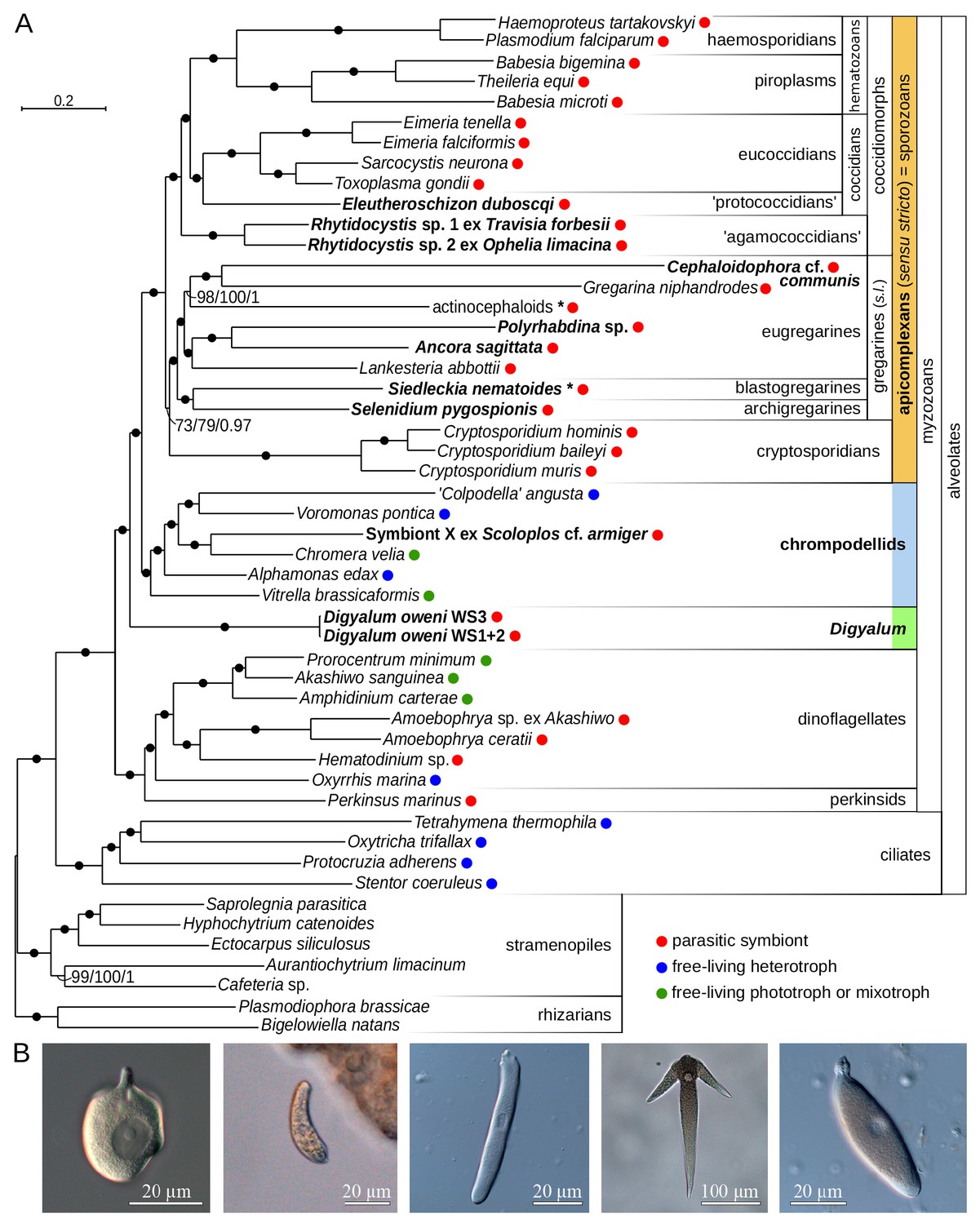
Synthetic tree reflecting the current phylogenetic framework of myzozoans (ciliates included as the outgroup). The tree shows that the Squirmida (Platyproteum spp., Filipodium, and Digyalum) branch separately from gregarines and form the sister lineage to a clade consisting of apicomplexans and chrompodellids. The myzozoan ancestor is inferred to have been a biflagellated, myzocytotic-feeding predator. Parasitism evolved independently multiple times within the Myzozoa, and flagella were lost in the most recent ancestor of apicomplexans in association with the origin of parasitism. The phylogenetic positions of the specific lineage investigated in this study, namely Platyproteum vivax, and the lineage of gregarine apicomplexans that most closely resembles Platyproteum, namely Selenidium, are written in bold.
| |||||||||||||||||||||
| Phylogeny of Squirmida according to Danja Currie-Olsen & Brian S. Leander (2024). |

Autor/Urheber: Danja Currie-Olsen & Brian S. Leander, Lizenz: CC BY 4.0
Confocal laser scanning micrographs (CLSM) of two different live trophozoites of Platyproteum vivax stained for mitochondria (green).
- (C, F) Superimposed Differential interference contrast (DIC) and mitochondria-stained CLSM images showing the dense layer of fluorescence sitting below the inner-membrane complex and plasma membrane.

Autor/Urheber: Danja Currie-Olsen & Brian S. Leander, Lizenz: CC BY 4.0
Scanning electron micrographs (SEM) of the trophozoites in Platyproteum vivax.
- (D, E) High magnification SEMs of the anterior apparatus showing the emergence of an anterior flagellum (AF) and a posterior flagellum (PF).
Autor/Urheber: Danja Currie-Olsen & Brian S. Leander, Lizenz: CC BY 4.0
Illustrations summarizing the overall organization of microtubules, basal bodies, and mitochondria in the trophozoites of Platyproteum vivax.
- (A) Dorsal and ventral views of a trophozoite showing the anterior apparatus With two flagella oriented to the right and left, respectively. Green denotes the position of the basal bodies from which the flagella are derived. Red denotes microtubules.
"Longitudinal microtubule bundles" (LMBs) are arranged in parallel along the entire anteroposterior axis of the cell, and
"dorsoventral microtubule bundles" (DVMBs), indicated by red dots, are arranged perpendicular to the anteroposterior axis of the cell. Most of the DVMBs are aligned in a distinctive curved row that runs from the anterior to the posterior end of the trophozoite. The row of DVMBs is positioned to the right of the nucleus in dorsal view and to the left of the nucleus in ventral view. A few isolated DVMBs are scattered on the side of the nucleus opposite of the curved row of DVMBs. - (B) Sagittal section showing the LMBs subtending the inner membrane complex and the perpendicular DVMBs running from the dorsal side to the ventral side of the trophozoite. Brown denotes the superficial layer of mitochondria located just above the LMB layer.
- (C) Cross sections through the nucleus of the trophozoite showing the LMBs subtending the inner membrane complex and a perpendicular DVMB running from the dorsal to ventral side of the cell.
Autor/Urheber: Danja Currie-Olsen & Brian S. Leander, Lizenz: CC BY 4.0
Scanning electron micrographs (SEM) of the trophozoites in Platyproteum vivax.
- (A—C) Trophozoites representing different stages of movement showing the anterior end oriented to the left, longitudinal surface folds (triple overhead arrows), transverse surface folds (arrowheads), and two relatively short flagella (double arrowheads).

Autor/Urheber: Danja Currie-Olsen & Brian S. Leander, Lizenz: CC BY 4.0
Confocal laser scanning micrographs (CLSM) of a trophozoite of Platyproteum vivax stained for alpha tubulin (red).
- (A) Differential interference contrast (DIC) light micrograph showing the dorsal surface of a trophozoite, nucleus (N), nucleolus (arrow), and anterior apparatus oriented upward and to the right.
- (B) Tubulin staining at a superficial focal plane showing the bundles of microtubules (red striations) running longitudinally along the entire length of the cell.
- (C) Superimposed Differential interference contrast (DIC) light micrograph and tubulin-stained Confocal laser scanning micrographs (CLSM) showing the longitudinal microtubule bundles positioned beneath the dorsal surface of the cell.

Autor/Urheber: Danja Currie-Olsen & Brian S. Leander, Lizenz: CC BY 4.0
Differential interference contrast (DIC) and confocal laser scanning micrographs (CLSM) showing two different trophozoites of Platyproteum vivax stained for centrin (green).
- (C) Superimposed DIC and centrin-stained CLSM images showing the position of the two distinct bodies (arrows) along the edge of the anterior apparatus and just below the plasma membrane; dorsal side of the cell.
- (F) Superimposed DIC and centrin-stained CLSM images showing the position of the two distinct bodies (arrows) along the edge of the anterior apparatus and just below the plasma membrane; ventral side of the cell.