Proteoarchaeota
| Proteoachaeota | ||||||
|---|---|---|---|---|---|---|
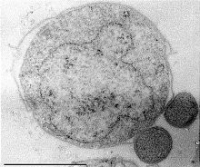
 Sulfolobus tengchongensis, | ||||||
| Systematik | ||||||
| ||||||
| Wissenschaftlicher Name | ||||||
| Proteoachaeota | ||||||
| Petitjean, Deschamps, López-García, Moreira, 2014 |
Proteoarchaeota (auch Proteoarchaea)[1] ist ein vorgeschlagenes Reich der Archaeen.
Systematik
Die phylogenetische Beziehung dieser Gruppe wird noch diskutiert.
- Einen vorgeschlagenen Hauptzweig bildet die „TACK-Supergruppe“ (Superphylum benannt nach den ersten Buchstaben der ihr zugeordneten „Gründungsmitglieder“ Thaum-, Aig-, Cren- und Korarchaeota) alias „Filarchaeota“.[2]
- Einen weiteren vorgeschlagenen Hauptzweig bildet die „Asgard-Supergruppe“ alias „Asgardarchaeota“[3] (Superphylum benannt nach Asgard, dem Reich der Götter in der nordischen Mythologie).
Die Beziehung der Mitglieder untereinander ist ungefähr wie folgt:[4][5][6][7][8][9]
| „Proteoarchaeota“ |
| |||||||||||||||||||||||||||||||||||||||||||||||||||||||||
Als Kandidaten für die Proto-Miochondrien unter den α-Proteobacteria wurden früher die Rickettsiales gehandelt, neuerdings (seit 2023) werden die zwischenzeitlich gefundenen und mit den Rickettsiales weitläufig verwandten Iodidimonadales favorisiert.
In einer alternativen Sicht stehen die Heimdallarchaeota den anderen „Asgard“-Archaeen (Lokiarchaeota etc.) näher als den Ur- bzw. Eukaryoten. Die Eukaryoten stehen dann außerhalb der „Asgard-Gruppe“ (als deren Schwester-Taxon) und bilden mit diesen zusammen ein Taxon „Eukaryomorpha“ (welches dann seinerseits das Schwestertaxon zur „TACK-Gruppe“ ist).[11][12]


Als weitere Mitgliederkandidaten wurden vorgeschlagen:
- „TACK-Superphylum“ („Filarchaeota“)
- „Asgard(archaeota)“
Siehe auch
- Eozyten-Hypothese (Eozyten: veraltet für Crenarchaeota)
- Letzter gemeinsamer Vorfahr der Eukaryoten (LECA)
- Histone – findet man bei Eukaryoten, Proteoarchaeota und Euryarchaeota
- Membranständige ATPasen (Homologiebeziehungen)
Literatur
- C. Petitjean, P. Deschamps, P. López-García, D. Moreira: Rooting the Domain archaea by phylogenomic analysis supports the foundation of the new kingdom proteoarchaeota. In: Genome Biol. Evol. 7. Jahrgang, Nr. 1, 2014, S. 191–204, doi:10.1093/gbe/evu274, PMID 25527841, PMC 4316627 (freier Volltext).
- Eugene V. Koonin: Archaeal ancestors of eukaryotes: not so elusive any more. In: BMC Biology. 13. Jahrgang, Nr. 1, 2015, S. 84, doi:10.1186/s12915-015-0194-5, PMID 26437773, PMC 4594999 (freier Volltext).
- John Timmer: The “Asgard archaea” are our own cells’ closest relatives, auf: Ars Technica, vom 13. Januar 2017
Einzelnachweise
- ↑ Jonathan Lombard: The multiple evolutionary origins of the eukaryotic N-glycosylation pathway, in: Biology Direct Band 11, Nr. 36, August 2016, doi:10.1186/s13062-016-0137-2
- ↑ T. Cavalier-Smith: The neomuran revolution and phagotrophic origin of eukaryotes and cilia in the light of intracellular coevolution and a revised tree of life. In: Cold Spring Harb. Perspect. Biol. 6. Jahrgang, Nr. 9, 2014, S. a016006, doi:10.1101/cshperspect.a016006, PMID 25183828, PMC 4142966 (freier Volltext).
- ↑ Paul-Adrian Bulzu, Adrian-Stefan Andrei, Michaela M. Salcher, Maliheh Mehrshad, Keiichi Inoue, Hideki Kandori, Oded Beja, Rohit Ghai, Horia L. Banciu: Casting light on Asgardarchaeota metabolism in a sunlit microoxic niche, in: Nat Microbiol, Band 4, S. 1129–1137, doi:10.1038/s41564-019-0404-y
- ↑ Anja Spang, Jimmy H. Saw, Steffen L. Jørgensen, Katarzyna Zaremba-Niedzwiedzka, Joran Martijn, Anders E. Lind, Roel van Eijk, Christa Schleper, Lionel Guy, Thijs J. G. Ettema: Complex archaea that bridge the gap between prokaryotes and eukaryotes. In: Nature. 521. Jahrgang, Nr. 7551, 2015, S. 173–179, doi:10.1038/nature14447, PMID 25945739, PMC 4444528 (freier Volltext), bibcode:2015Natur.521..173S (englisch).
- ↑ Katarzyna Zaremba-Niedzwiedzka, Eva F. Caceres, Jimmy H. Saw, Disa Bäckström, Lina Juzokaite, Emmelien Vancaester, Kiley W. Seitz, Karthik Anantharaman, Piotr Starnawski, Kasper U. Kjeldsen, Matthew B. Stott, Takuro Nunoura, Jillian F. Banfield, Andreas Schramm, Brett J. Baker, Anja Spang, Thijs J. G. Ettema: Asgard archaea illuminate the origin of eukaryotic cellular complexity. In: Nature. 541. Jahrgang, Nr. 7637, 2017, S. 353–358, doi:10.1038/nature21031, PMID 28077874, bibcode:2017Natur.541..353Z.
- ↑ Katarzyna Zaremba-Niedzwiedzka et al.: Asgard archaea illuminate the origin of eukaryotic cellular complexity. Nature 541, 19. Januar 2017, S. 353–358, doi:10.1038/nature21031
- ↑ Gregory P. Fournier, Anthony M. Poole: A Briefly Argued Case That Asgard Archaea Are Part of the Eukaryote Tree. In: Frontiers in Microbiology. 9. Jahrgang, 2018, ISSN 1664-302X, S. 1896, doi:10.3389/fmicb.2018.01896, PMID 30158917, PMC 6104171 (freier Volltext) – (englisch).
- ↑ M. P. Ferla, J. C. Thrash, S. J. Giovannoni, W. M. Patrick: New rRNA gene-based phylogenies of the Alphaproteobacteria provide perspective on major groups, mitochondrial ancestry and phylogenetic instability. In: PLoS ONE. 8. Jahrgang, Nr. 12, 2013, S. e83383, doi:10.1371/journal.pone.0083383, PMID 24349502, PMC 3859672 (freier Volltext), bibcode:2013PLoSO...883383F.
- ↑ Laura Eme, Anja Spang, Jonathan Lombard, Courtney W. Stairs, Thijs J. G. Ettema: Archaea and the origin of eukaryotes. In: Nature Reviews Microbiology. 15. Jahrgang, Nr. 12, 10. November 2017, ISSN 1740-1534, S. 711–723, doi:10.1038/nrmicro.2017.133, PMID 29123225 (englisch).
- ↑ Céline Brochier-Armanet, Bastien Boussau, Simonetta Gribaldo, Patrick Forterre: Mesophilic crenarchaeota: Proposal for a third archaeal phylum, the Thaumarchaeota. In: Nature Reviews Microbiology. Band 6, Nr. 3, 2008, S. 245–252, doi:10.1038/nrmicro1852, PMID 18274537.
- ↑ Gregory P. Fournier, Anthony M. Poole: A Briefly Argued Case That Asgard Archaea Are Part of the Eukaryote Tree. In: Frontiers in Microbiology. 9. Jahrgang, 15. August 2018, ISSN 1664-302X, doi:10.3389/fmicb.2018.01896, PMID 30158917, PMC 6104171 (freier Volltext).
- ↑ Nicholas P Robinson: Archaea, from obscurity to superhero microbes: 40 years of surprises and critical biological insights, auf: ResearchGate vom Dezember 2018, doi:10.1042/ETLS20180022, Fig. 1
- ↑ a b Yinzhao Wang, Gunter Wegener, Jialin Hou, Fengping Wang, Xiang Xiao. Expanding anaerobic alkane metabolism in the domain of Archaea, in: Nature Microbiologyvolume 4, S. 595–602, vom 4. März 2019, doi:10.1038/s41564-019-0364-2
- ↑ a b Fengping Wang:Behind the paper: Expanding anaerobic alkane metabolism in the domain of Archaea ( des vom 22. April 2019 im Internet Archive) Info: Der Archivlink wurde automatisch eingesetzt und noch nicht geprüft. Bitte prüfe Original- und Archivlink gemäß Anleitung und entferne dann diesen Hinweis., in: Nature Microbiology (Contributor) vom 4. März 2019
- ↑ a b I. Vanwonterghem, P. N. Evans, D. H. Parks, P. D. Jensen, B. J. Woodcroft, P. Hugenholtz, G. W. Tyson: Methylotrophic methanogenesis discovered in the archaeal phylum Verstraetearchaeota, in: Nat Microbiol.; 1:16170., vom 3. Oktober 2016, doi:10.1038/nmicrobiol.2016.170, PMID 27694807
- ↑ Kiley W. Seitz, Nina Dombrowski, Laura Eme, Anja Spang, Jonathan Lombard, Jessica R. Sieber, Andreas P. Teske, Thijs J. G. Ettema, Brett J Baker: Asgard archaea capable of anaerobic hydrocarbon cycling, in: Nature Communications, Dezember 2019 doi:10.1038/s41467-019-09364-x
Auf dieser Seite verwendete Medien
Autor/Urheber: (of code) -xfi-, Lizenz: CC BY-SA 3.0
The Wikispecies logo created by Zephram Stark based on a concept design by Jeremykemp.
Autor/Urheber: Hiroyuki Imachi, Masaru K. Nobu, Nozomi Nakahara, Yuki Morono, Miyuki Ogawara, Yoshihiro Takaki, Yoshinori Takano, Katsuyuki Uematsu, Tetsuro Ikuta, Motoo Ito, Yohei Matsui, Masayuki Miyazaki, Kazuyoshi Murata, Yumi Saito, Sanae Sakai, Chihong Song, Eiji Tasumi, Yuko Yamanaka, Takashi Yamaguchi, Yoichi Kamagata, Hideyuki Tamaki, Ken Takai, Lizenz: CC BY-SA 4.0
English: Ca. Prometheoarchaeum syntrophicum aka Lokiarchaeota sp. MK-D1. SEM images of MK-D1 cells producing long branching (g) and straight (h) membrane protrusions. Scale bars, 1 μm.
Bild über den Reitenden Urzwerg
Autor/Urheber: Jong-Geol Kim, Khaled S. Gazi, Mart Krupovic, Sung-Keun Rhee, Lizenz: CC BY-SA 4.0
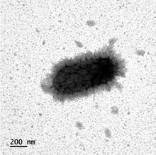
Thaspiviridae, species Nitmarvirus NSV1. Nitrosopumilus spindle-shaped virus 1 particles attached to the surface of a host cell. Scale bar, 200 nm
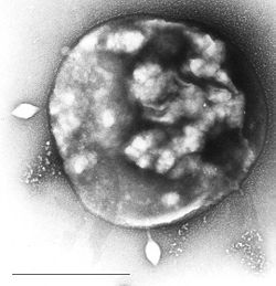
Cell of Sulfolobus infected by virus STSV1 observed under microscopy. Two spindle-shaped viruses were being released from the host cell. The strain of Sulfolobus and STSV1 (Sulfolobus tengchongensis Spindle-shaped Virus 1) were isolated by Xiaoyu Xiang and his colleagues in an acidic hot spring in Yunnan Province, China. At present, STSV1 is the largest archaeal virus to have been isolated and studied. Its genome sequence has been sequenced.


