Typisches Aussehen eines Virusteilchens mit Myoviren-Morphologie, sheath: Schwanzscheide, baseplate: BasisplatteDetailzeichnung im Fall eines Bakteriophage vom Morphotyp der Myoviren mit kontraktilem Schwanzeil
Die morphologisch begründete (nicht-taxonomische) Gruppe der Myoviren (englischmyoviruses, früher auch Morphotyp A genannt) umfasst eine Reihe von Familien, Unterfamilien und Gattungen von Viren mit einem linearen Molekül doppelsträngiger DNA (dsDNA) als Genom.
Die Myoviren werden unterteilt in Subtypen: 1: Kopf ohne „Fühler“, aber kurze Anhängsel am Schwanz; 2: kragenartige Struktur zwischen Kopf und Schwanz und kurze Anhängsel am Schwanz.[2]
Alle Mitglieder gehören zur Klasse der Caudoviricetes („Schwanzviren“ – Viren mit Kopf-Schwanz-Struktur): Ihre Mitglieder besitzen ein ikosaedrischesKapsid und ein Schwanzteil; wegen dessen kontraktiler Eigenschaft leitet sich der Gruppenname von griechischμυόςmyos, deutsch ‚Muskel‘ ab. Als Wirte dienen Prokaryoten, meist Bakterien (Bakteriophagen), aber teilweise auch Archaeen. Innerhalb der Klasse Caudoviricetes zeichnen sich die Myoviren außerdem durch besonders große, teilweise langgestreckte Kapside (z. B. T4-Phage: 111 × 78 nm) und ein sehr großes Genom (34–169 kBp) aus (siehe Riesenphagen). Der wichtigste Vertreter ist der schon früh entdeckte T4-Phage (Spezies Enterobacteria-Virus T4, Gattung Tequatrovirus), dessen besondere Morphologie und genetische Eigenschaften in der molekularbiologischen Forschung eine herausragende Rolle spielte. Die von diesem Phagen abgeleitete T4-DNA-Ligase findet heute noch Verwendung in der Molekularbiologie. Weitere Mitglieder von Bedeutung sind der Vibrio-Phage KVP40 (Spezies Vibrio-Virus KVP40, Gattung Schizotequatrovirus), der Pseudomonas-Phage phiKZ (Spezies Pseudomonas-Virus phiKZ, Gattung Phikzvirus), der Bakteriophage P1 (Spezies Escherichia-Virus P1, Gattung Punavirus, siehe P1 Artificial Chromosome) u. a.
Die Gruppe galt lange Zeit als ein Virustaxon im Rang einer Virusfamilie mit der Bezeichnung Myoviridae. Im März 2021 wurde vorgeschlagen, diese Familie mitsamt der Ordnung Caudovirales wegen fehlender Monophylie aufzulösen und (wie damals bereits z. T. geschehen) durch neu zu schaffende Familien zu ersetzen, damit neue Ergebnisse aus der Metagenomik in die Taxonomie aufgenommen werden können.[3] Das International Committee on Taxonomy of Viruses (ICTV) hat dem im März 2022 entsprochen.[1] Gemäß Vorschlag bleibt die Bezeichnung „Myoviren“ (englischmyoviruses) aber als informeller Sammelbegriff morphologisch ähnlicher Prokaryotenviren mit einem linearen Doppelstrang-DNA-Genom erhalten.[3]
Listeria-Phage A511 (Herelleviridae): Aufbau der Basisplatte und Veränderung während der Absorption an der Zellwand des Wirtsbalterium.
Spezies „Campylobacter virus IBB35“ („Campylobacter-Virus IBB35“), mit Campylobacter-Phage vB_CcoM-IBB_35[19]
Schemazeichnung eines Virions der Gattung Fletchervirus (Campylobacter-Phage CP81) mit Schwanzfibern. Bei der Schwestergattung Firehammervirus (Campylobacter-Phage CP220) sind diese nicht sicher vorhanden, vgl. Zeichnung oben Ackermannviridae
Spezies „Mycobacterium-Phage Nidhogg“ (en. „Mycobacterium phage Nidhogg“) (Vorschlag, wohl zu unterscheiden von Asgardviren mit vorgeschlagener Bezeichnung Nidhogg-Viren, en. Nidhogg viruses)[21]
Spezies „…“ (weitere Vorschläge)
Gattung Myrnavirus
Spezies Myrnavirus myrna (Mycobacterium-Virus Myrna), mit Mycobacterium-Phage Myrna
Spezies Myrnavirus phabba, mit Mycobacterium-Phage Phabba
Unterfamilie Iiscvirinae
Gattung Aryavirus
Spezies Aryavirus arya, mit Enterobacter-Phage Arya
Gattung Jilinvirus (früher Cvm10virus, zu ehem. Familie Myoviridae)[22]
Spezies Jilinvirus ECOO78, mit Escherichia phage vB_EcoM_ECOO78
Spezies Jilinvirus ep3, mit Escherichia phage vB_EcoM-ep3
Unterfamilie Jameshumphriesvirinae
Gattung Bimevirus (abgetrennt von Jedunavirus)
Spezies Bimevirus bv1611EK21
Spezies Bimevirus IME346, mit Klebsiella-Phage vB_KpnM_IME346
Spezies Bimevirus KB2
Gattung Chaoyangvirus
Spezies Chaoyangvirus BUCT49532, mit Klebsiella-Phage BUCT_49532
Gattung Geezettvirus
Spezies Geezettvirus geezett, mit Klebsiella-Phage Geezett
Gattung Jedunavirus, früher zur ehem. Familie Myoviridae[23]
Spezies Jedunavirus JD001, mit Klebsiella-Phage JD001
Gattung Mascletvirus
Spezies Mascletvirus VLCpiM12a, mit Klebsiella-Phage VLCpiM12a
Gattung Parissaclayvirus
Spezies Parissaclayvirus POU148, mit Klebsiella-Phage vB_KpnM_15-38_KLPPOU148
Gattung Peatvirus
Spezies Peatvirus peat2, mit Pectinobacterium-Phage PEAT2
Gattung Ringroadvirus
Spezies Ringroadvirus BUCT47333, mit Klebsiella-Phage BUCT_47333
Gattung Sircambvirus
Spezies Sircambvirus FZ14, mit Klebsiella-Phage vB_KpnM_FZ14
Spezies Sircambvirus justaphage, mit Klebsiella-Phage vB_KpnM_JustaPhage
Spezies Sircambvirus KpV52, mit Klebsiella-Phage vB_KpnM_KpV52
Spezies Sircambvirus KpV79, mit Klebsiella-Phage vB_KpnM_KpV79
Spezies Sircambvirus MEW1, mit Klebsiella-Phage MEW1
Spezies Sircambvirus pKp383, mit Klebsiella-Phage pKp383
Gattung Zewailvirus
Spezies Zewailvirus SBP, mit Klebsiella-Phage SBP
Spezies Zewailvirus ZCEC13, mit Escherichia-Phage ZCEC13
Unterfamilie Kantovirinae
Gattung Beograduvirus
Spezies Beograduvirus KPhi1
Spezies Beograduvirus Xaf18
Gattung Tsukubavirus
Spezies Tsukubavirus OP2 (Xanthomonas-Virus OP2, früher in Gattung Naesvirus)
Spezies Tsukubavirus XPP1
Spezies Tsukubavirus XPV1
Unterfamilie Stephanstirmvirinae
Gattung Justusliebigvirus
Spezies Justusliebigvirus alia, mit Escherichia-Phage alia
Spezies Justusliebigvirus muut, mit Escherichi-Phage muut
Spezies Justusliebigvirus PD06, mit Escherichia-Phage vB_vPM_PD06
Spezies Justusliebigvirus PHB05, mit Escherichia-Phage vB_EcoM_PHB05
Spezies Justusliebigvirus phi92, mit Enterobacteria-Phage phi92[24][25]
Spezies Justusliebigvirus VEcB, mit Escherichia-Phage VEcB
Gattung Phapecoctavirus (13 Spezies)
Spezies Phapecoctavirus anhysbys, mit Escherichia-Phage anhysbys
Spezies Muvirus mu (Escherichia-Virus Mu), mit Enterobacteria-Phage Mu, alias Bacteriophage Mu oder Myovirus Mu[45] (siehe Barbara McClintock §Nobelpreis)
Spezies Muvirus SfMu (Shigella-Virus SfMu)
TEM-Aufnahme zweier Virionen von Alteromonas-Phage AltPT11-V22, Gattung Myoalterovirus.
Gattung Myoalterovirus
Spezies Myoalterovirus PT11V22 (Alteromonas-Myovirus V22, Alteromonas-Virus AltPT11-V22), mit Alteromonas-Phage AltPT11-V22 alias Phage vB_AmeM_PT11-V22[46]
Spezies Shirahamavirus PTm1 (Tenacibaculum-Virus PTm1), mit Tenacibaculum-Phage PTM1 alias PTm1 und Tenacibaculum-Phage PTm5[53][40] – verwandt mit Tenacibaculum-Virus pT24 (Kungbxnavirus) und Sphingomonas-Phage PAU[40]
Gattung Svunavirus
Spezies Svunavirus GBSV1, mit Geobacillus-Phage GBSV1
Spezies Svunavirus sv1 (Bacillus-Virus 1), mit Bacillus-Phage 1 (DQ840344)[A. 2]
Gattung Takahashivirus
Spezies Takahashivirus PBS1 (Bacillus-Virus PBS1), mit Bacillus-Phage PBS1 alias Bacillus subtilis phage PBS1[55][56][57]
Spezies „Sphingomonas-Phage PAU“[77][78] – verwandt mit Tenacibaculum-Virus pT24 (Kungbxnavirus) und Tenacibaculum-Virus PTm1 (Shirahamavirus),[40]Jumbo-Phage mit einer Mopp-ähnlichen Anordnung von flexiblen Schwanzfasern[7]
Spezies „Bacillus-Phage PBS2“ (en. „Bacillus phage PBS2“) mit Bacteriophage PBS2.[79]
Spezies „Alishewanella phage vB_AspM_Slicko01“ mit Phage Slicko[83][85]
Spezies „Vibrio phage 24“ mit Vibrio cholerae phage 24[86][87]
Spezies „Vibrio phage X29“ mit Vibrio cholerae phage X29[88][43][87]
Riesenphagen
→ Hauptartikel: Riesenphagen
Die am 3. März 2025 vom ICTV als Mitglieder der neuen Ordnung Grandevirales offiziell anerkannten Lak-Phagen wurden von Audra E. Devoto, Jillian F. Banfieldet al. im Januar 2019 erstbeschrieben. Aufgrund ihrer Genomgröße von mehr als 200 kbp (Kilobasenpaaren), darunter ein Genom von 735 kbp (das bis dato größte beschriebene Phagengenom) werden diese als ‚Riesenphagen‘ klassifiziert. Das Team hatte das Darm-Mikrobiom von Personen mit nicht-westlicher Ernährungsweise aus der Verwaltungseinheit (Upazila) Laksam in der Division Chittagong (Bangladesch) und der Hadza aus Tansania, sowie zum Vergleich auch von kenianischenGelben Pavianen (Papio cynocephalus) und Dänischen Schweinen untersucht. Wie sich zeigt, infizierten die gefundenen Riesenphagen Darmbakterien der Gattung Prevotella. Nach den Ergebnissen sind Lak-Phagen häufige und offenbar wichtige Bestandteile des Darmmikrobioms von Menschen und Tieren. Es wurde zudem festgestellt, dass von den Lak-Phagen das kanonische TAG-Stoppcodon (alias UAG-Stoppcodon, Uracil–Adenin–Guanin) offenbar zur Codierung der AminosäureGlutamin (Q, Code 15) umfunktioniert wird.[8] Weitere Untersuchungen von Ryan Cook, Evelien M. Adriaenssens et al. (2024) führten dann zum 2025 vom ICTV bestätigten Vorschlag der neuen Ordnung, Grandevirales, die sich insbesondere durch eine Uminterpretation des UAG-Codons zwecks Codierung einer Aminosäure auszeichnet.[10]
Zwischenzeitlich hatten Untersuchungen von Basem Al-Shayeb, Jillian F. Banfieldet al. im Februar 2020 weitere Kladen von Riesenpagen identifiziert, die von dem Team mit den provisorischen Bezeichnungen „Kabirphage“, „Mahaphage“, „Biggiephage“, „Dakhmphage“, „Kyodaiphage“, „Kaempephage“, „Jabbarphage“, „Enormephage“, „Judaphage“ und „Whopperphage“ (alles verschiedene Bezeichnungen für „groß“, „riesig“) benannt wurden. Einige dieser neu entdeckten Riesenphagen tragen Gene für Varianten der Cas-Proteine, die in verschiedenen bakteriellen CRISPR-Systemen zu finden sind, z. B. in den FamilienCas9, Cas12, CasX und CasY. Die Lak-Phagen erwiesen sich zudem als Teil der „Mahaphage“-Klade,[89] sodass eine Synonymie dieser Klade mit der inzwischen eingerichteten Ordnung Grandevirales nahe liegt.
Bacillus-Phage 1_ICo-2020 (MT700412, Spezies Suttonboningtonvirus sv1ICo2020, Familie Ehrlichviridae)
„Streptomyces-Phage SV1“ (ΦSV1, JX182371 – vorschlagsgemäß zur Gattung Picardvirus),[54]
Literatur
A. M. Q. King, M. J. Adams, E. B. Carstens, E. J. Lefkowitz (Hrsg.): Virus Taxonomy. Ninth Report of the International Committee on Taxonomy of Viruses. Amsterdam 2012, ISBN 978-0-12-384684-6, S. 46–62.
C. M. Fauquet, M. A. Mayo et al.: Eighth Report of the International Committee on Taxonomy of Viruses. London / San Diego 2004.
C. M. Mizuno, F. Rodriguez-Valera, N. E. Kimes, R. Ghai: Expanding the Marine Virosphere Using Metagenomics. In: PLoS Genetics. 9. Jahrgang, Nr.12, 2013, S.e1003987, doi:10.1371/journal.pgen.1003987, PMID 24348267, PMC 3861242 (freier Volltext) – (englisch).
↑ Antje Wichels, Stefan S. Biel, Hans R. Gelderblom, Thorsten Brinkhoff, Gerard Muyzer, Christian Schütt: Bacteriophage diversity in the North Sea. In: Applied and Environmental Microbiology, Band 64, Nr. 11, November 1998, S. 4128-4133; doi:10.1128/AEM.64.11.4128-4133.1998, PMID 9797256, PMC 106618 (freier Volltext), PDF (englisch).
↑ ab Dann Turner, Andrew M. Kropinski, Evelien M. Adriaenssens: A Roadmap for Genome-Based Phage Taxonomy. In: MDPI Viruses, Band 13, Nr. 3, Section Bacterial Viruses, 18. März 2021, S. 506; doi:10.3390/v13030506 (englisch).
↑ abcdefgh Lakshminarayan M. Iyer, Vivek Anantharaman, Arunkumar Krishnan, A. Maxwell Burroughs, L. Aravind: Jumbo Phages: A Comparative Genomic Overview of Core Functions and Adaptions for Biological Conflicts. In: MDPI: Viruses, Band 13, Nr. 1, 5. Januar 2021, S. 63; doi:10.3390/v13010063 (englisch).
↑ ab Audra E. Devoto, Joanne M. Santini et al.: Megaphages infect Prevotella and variants are widespread in gut microbiomes. In: Nature Microbiology, Band 4, 28. Januar 2019, S. 693–700; doi:10.1038/s41564-018-0338-9 (englisch). Siehe insbes. Tabelle 1 und Supplementary Figure 11.
↑ Igor Babkin, Artem Tikunov, Vera Morozova, Andrey Matveev, Vitaliy V. Morozov, Nina Tikunova: Genomes of a Novel Group of Phages That Use Alternative Genetic Code Found in Human Gut Viromes. In: MDPI: International Journal of Molecular Sciences, Band 24, Nr. 20, 18. Oktober 2023, S. 15302; doi:10.3390/ijms242015302, PMC 10607447 (freier Volltext), PMID 37894982 (englisch).
↑ ab Ryan Cook, Marco A. Crisci, Hannah V. Pye, Andrea Telatin, Evelien M. Adriaenssens, Joanne M. Santini: Decoding huge phage diversity: a taxonomic classification of Lak megaphages Open Access. In: Journal of General Virology Band 105, Nr. 5, 30. Mai 2024; doi:10.1099/jgv.0.001997 (englisch). Dazu:
Ryan Cook, Hannah V. Pye, M. A. Crisci, J. M. Santini, Eveleian M. Adriaenssens: Create one new order Grandevirales (Duplodnaviria).Proposal 2024.014B (zip:docx). Vorschlag an das ICTV vom 4. Juni 2023 mit Revision vom 7. Oktober 2024 (angenommen).
↑ Y. Liu et al. (ICTV Archaeal Viruses Subcommittee): Proposal 2021.001A (zip:docx), PDF (via Universität Helsinki). Create three new orders and 14 new families in the class Caudoviricetes (Duplodnaviria, Uroviricota) for classification of archaeal tailed viruses. Oktober 2020. Siehe insbes. Tbl. 1.
↑ C. Lood, R. Lavigne, D. Turner, C. Moraru, E. M. Adriaenssens, A. M. Kropinski, Z. Drulis-Kawa: Proposal 2022.006B Create a new family (Arenbergviridae) and a new genus (Wroclawvirus) with a single species (Caudoviricetes). Oktober 2020.
↑ Enea Maffei, Anne-Kathrin Woischnig, Marco R. Burkolter, Yannik Heyer, Dorentina Humolli, Nicole Thürkauf, Thomas Bock, Alexander Schmidt, Pablo Manfredi, Adrian Egli, Nina Khanna, Urs Jenal, Alexander Harms: Phage Paride can kill dormant, antibiotic-tolerant cells of Pseudomonas aeruginosa by direct lytic replication. In: Nature Communications, Band 15, Nr. 175, 2. Januar 2024; doi:10.1038/s41467-023-44157-3 (englisch). Dazu:
↑ ab L. Truncaite, E. Šimoliūnas, A. Zajančkauskaite, L. Kaliniene, R. Mankevičiūte, J. Staniulis, V. Klausa, R. Meškys: Bacteriophage vB_EcoM_FV3: A new member of "rV5-like viruses". In: Archives of Virology. 157. Jahrgang, Nr.12, 2012, S.2431–2435, doi:10.1007/s00705-012-1449-x, PMID 22907825 (englisch).
↑ S. B. Santos, A. M. Kropinski, P.-J. Ceyssens, H.-W. Ackermann, A. Villegas, R. Lavigne, V. N. Krylov, C. M. Carvalho, E. C. Ferreira, J. Azeredo: Genomic and Proteomic Characterization of the Broad-Host-Range Salmonella Phage PVP-SE1: Creation of a New Phage Genus. In: Journal of Virology. 85. Jahrgang, Nr.21, 2011, S.11265–11273, doi:10.1128/JVI.01769-10, PMID 21865376, PMC 3194984 (freier Volltext) – (englisch).
↑ abcdefgh André M. Comeau, Denise Tremblay, Sylvain Moineau, Thomas Rattei, Alla I. Kushkina, Fedor I. Tovkach, Henry M. Krisch, Hans-Wolfgang Ackermann: Phage Morphology Recapitulates Phylogeny: The Comparative Genomics of a New Group of Myoviruses. In: PLOS ONE, Band 7, Nr. 7, 6. Juli 2012, S. e40102; doi:10.1371/journal.pone.0040102, PMID 22792219 (englisch).
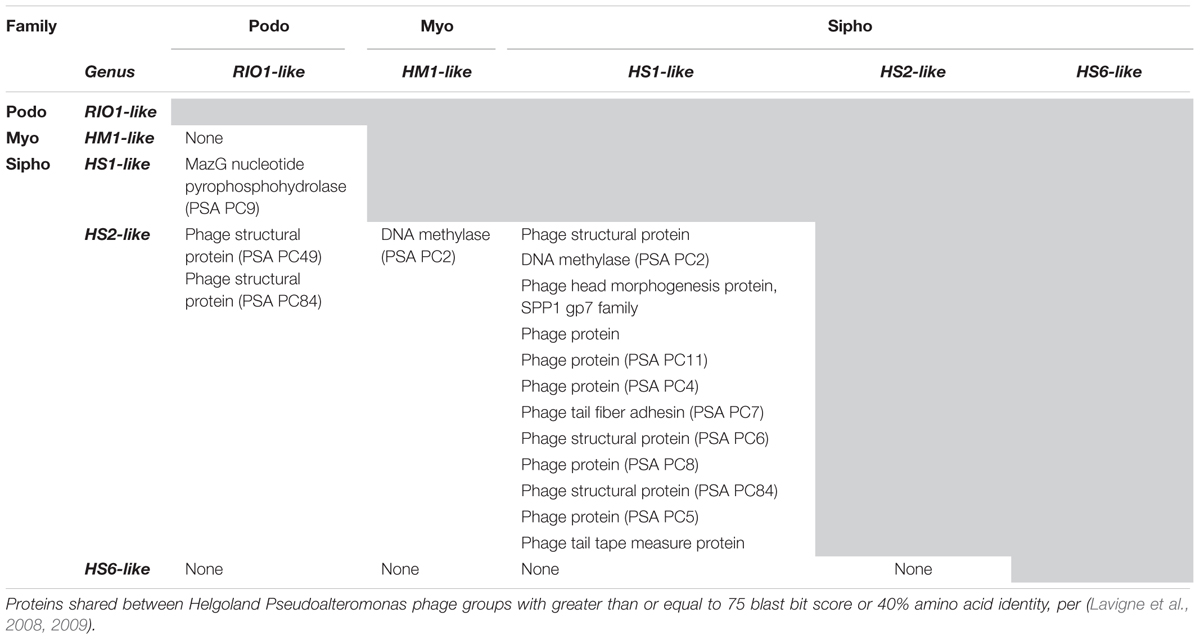
↑ Melissa B. Duhaime, Natalie Solonenko, Simon Roux, Nathan C. Verberkmoes, Antje Wichels, Matthew B. Sullivan: Comparative Omics and Trait Analyses of Marine Pseudoalteromonas Phages Advance the Phage OTU Concept. In: Frontiers in Microbiology, Band 8, Sec. Virology, 6. Juli 2017, S. 1241; doi:10.3389/fmicb.2017.01241, PMID 28729861, PMC 5498523 (freier Volltext) (englisch). Siehe insbes. Tbl. 2.
↑ Grégory Resch, Eva M. Kulik, Fred S. Dietrich, Jürg Meyer: Complete Genomic Nucleotide Sequence of the Temperate Bacteriophage AaΦ23 of Actinobacillus actinomycetemcomitans. In: ASM Journals: Journal of Bacteriology, Band 186, Nr. 16, 15. August 2004, S. 5523–5528; doi:10.1128/JB.186.16.5523-5528.2004, PMC 490939 (freier Volltext), PMID 15292156 (englisch).
↑Eunsu Ha, Bokyung Son, Sangryeol Ryu: Clostridium perfringens Virulent Bacteriophage CPS2 and Its Thermostable Endolysin LysCPS2. In: MDPI. Viruses, Band 10, Nr. 5, Special Issue Biotechnological Applications of Phage and Phage-Derived Proteins, 251, 11. Mai 2018; doi:10.3390/v10050251 (englisch).
↑ Marcel Sprenger, Malte Siemers, Sebastian Krautwurst, Kai Papenfort: Small RNAs direct attack and defense mechanisms in a quorum sensing phage and its host. In: Cell Host & Microbe, 4. April 2024; doi:10.1016/j.chom.2024.03.010 (englisch). Dazu:
↑Katherine S. Wetzel, Haley G. Aull, Kira M. Zack, Rebecca A. Garlena, Graham F. Hatfull: Protein-Mediated and RNA-Based Origins of Replication of Extrachromosomal Mycobacterial Prophages. In: mBio, Band 11, Nr. 2, März/April 2020, e00385-20; doi:10.1128/mBio.00385-20, PMC 7157519 (freier Volltext), PMID 32209683, Epub 24. März 2020.
↑ K. Holmfeldt, N. Solonenko, M. Shah, K. Corrier, L. Riemann, N. C. Verberkmoes, M. B. Sullivan: Twelve previously unknown phage genera are ubiquitous in global oceans. In: Proceedings of the National Academy of Sciences. 110. Jahrgang, Nr.31, 2013, S.12798–12803, doi:10.1073/pnas.1305956110, PMID 23858439, PMC 3732932 (freier Volltext) – (englisch). Siehe insbes. Supplement 1.
↑ abC. Chénard, J. F. Wirth, C. A. Suttle: Viruses Infecting a freshwater filamentous cyanobacterium (Nostoc sp.) encode a functional CRISPR array and a proteobacterial DNA polymerase B. In: mBio. 7. Jahrgang, 14. Juni 2016, S.e00667–16, doi:10.1128/mBio.00667-16, PMID 27302758, PMC 4916379 (freier Volltext) – (englisch).
↑Andrea C. Baker, Victoria J. Goddard, Joanne Davy, Declan C. Schroeder, David G. Adams, William H. Wilson: Identification of a Diagnostic Marker To Detect Freshwater Cyanophages of Filamentous Cyanobacteria. In: ASM Journals. Applied and Environmental Microbiology, Band 72, Nr. 9, 6. September 2006; doi:10.1128/AEM.00270-06, PMID 16957185, PMC 1563665 (freier Volltext) (englisch).
↑ Ulrike Pfreundt, Dina Spungin, Shengwei Hou, Björn Voß, Ilana Berman-Frank, Wolfgang R. Hess: Genome of a giant bacteriophage from a decaying Trichodesmium bloom. In: Marine Genomics, Band 33, Juni 2017, S. 21-25; doi:10.1016/j.margen.2017.02.001, 314087529 (englisch).
↑Przemyslaw Decewicz, Piotr Golec, Mateusz Szymczak, Monika Radlinska, Lukasz Dziewit: Identification and Characterization of the First Virulent Phages, Including a Novel Jumbo Virus, Infecting Ochrobactrum spp. In: MDPI Int. J. Mol. Sci, Band 21, Nr. 6, Special Issue Bacteriophage Molecular Studies, 18. März 2020, 2096; doi:10.3390/ijms21062096 (englisch).
↑ A. Cornelissen, S. C. Hardies, O. V. Shaburova, V. N. Krylov, W. Mattheus, A. M. Kropinski, R. Lavigne: Complete Genome Sequence of the Giant Virus OBP and Comparative Genome Analysis of the Diverse KZ-Related Phages. In: Journal of Virology. 86. Jahrgang, Nr.3, 2011, S.1844–1852, doi:10.1128/JVI.06330-11, PMID 22130535, PMC 3264338 (freier Volltext) – (englisch).
↑ Richard Allen White III, Curtis A. Suttle: The Draft Genome Sequence of Sphingomonas paucimobilis Strain HER1398 (Proteobacteria), Host to the Giant PAU Phage, Indicates That It Is a Member of the Genus Sphingobacterium (Bacteroidetes). In: Genome Announcements. Band 1, Nr. 4, Juli-August 2013, Artikel e00598-13; doi:10.1128/genomeA.00598-13, PMC 3738902 (freier Volltext), PMID 23929486 (englisch).
↑Natalia Bagińska, Anna Pichlak, Andrzej Górski1, Ewa Jończyk-Matysiak: Specific and Selective Bacteriophages in the Fight against Multidrug-resistant Acinetobacter baumannii. In: Virologica Sinica, Band 34, S. 347–357; doi:10.1007/s12250-019-00125-0 (englisch): Siehe insbes. Tbl. 1.
↑Kaitlyn E. Kortright, Rachel E. Done, Benjamin K. Chan, Valeria Souza, Paul E. Turner: Selection for phage resistance reduces virulence of Shigella flexneri. In: ASM Appl. and Env. Microbiol. (AEM), 17. November 2021; doi:10.1128/AEM.01514-21 (englisch). Dazu:
↑ ab Janina Rahlff, Matthias Wietz, Helge-Ansgar Giebel, Oliver Bayfield, Emelie Nilsson, Kristofer Bergström, Kristopher Kieft, Karthik Anantharaman, Mariana Ribas-Ribas, Hannah D. Schweitzer, Oliver Wurl, Matthias Hoetzinger, Alfred Antson, Karin Holmfeldt: Ecogenomics and cultivation reveal distinctive viral-bacterial communities in the surface microlayer of a Baltic Sea slick. In: Oxford Academic: ISME Communications, Band 3, Nr. 1, Dezember 2023, S. 97; doi:10.1038/s43705-023-00307-8 (englisch).
↑ ab Sudhakar G. Bhandare, Andrew Warry, Richard D. Emes, Steven P. T. Hooton, Paul A. Barrow, Robert J. Atterbury: Complete Genome Sequences of Vibrio cholerae-Specific Bacteriophages 24 and X29. In: Genome Announcements, Band 5, Nr. 46, 16. November 2017, S. e01013-17; doi:10.1128/genomeA.01013-17, PMC 5690320 (freier Volltext), PMID 29146843 (englisch).
↑ Basem Al-Shayeb, Rohan Sachdeva, Lin-Xing Chen, Fred Ward, Patrick Munk, Audra Devoto, Cindy J. Castelle, Matthew R. Olm, Keith Bouma-Gregson, Yuki Amano, Christine He, Raphaël Méheust, Brandon Brooks, Alex Thomas, Adi Lavy, Paula Matheus-Carnevali, Christine Sun, Daniela S. A. Goltsman, Mikayla A. Borton, Jillian F. Banfieldet al.: Clades of huge phages from across Earth’s ecosystems. In: Nature, Band 578, 12. Februar 2020, S. 425–431; doi:10.1038/s41586-020-2007-4 (englisch). Dazu:
43705 2023 307 fig8a Slickus.jpg Autor/Urheber: Janina Rahlff, Matthias Wietz, Helge-Ansgar Giebel, Oliver Bayfield, Emelie Nilsson, Kristofer Bergström, Kristopher Kieft, Karthik Anantharaman, Mariana Ribas-Ribas, Hannah D. Schweitzer, Oliver Wurl, Matthias Hoetzinger, Alfred Antson, Karin Holmfeldt,
Lizenz:CC BY 4.0 Negatively stained electron micrographs reveal myovirus morphology of phage Slickus, scale bar 100 nm. Coverage of reads based on mapping to the host MAG Alishewanella sp. (MAG_01) as well as phage vB_AspM_Slickus01
43705 2023 307 fig8a Slicko.jpg Autor/Urheber: Janina Rahlff, Matthias Wietz, Helge-Ansgar Giebel, Oliver Bayfield, Emelie Nilsson, Kristofer Bergström, Kristopher Kieft, Karthik Anantharaman, Mariana Ribas-Ribas, Hannah D. Schweitzer, Oliver Wurl, Matthias Hoetzinger, Alfred Antson, Karin Holmfeldt,
Lizenz:CC BY 4.0 Negatively stained electron micrographs reveal myovirus morphology of phage Slicko, scale bar 100 nm. Coverage of reads based on mapping to the host MAG Alishewanella sp. (MAG_01) as well as phage vB_AspM_Slicko01
Structure of a Myoviridae bacteriophage 2.jpg Autor/Urheber: Chelsea Bonnain, Mya Breitbart and Kristen N. Buck,
Lizenz:CC BY-SA 4.0 Structure of a typical bacteriophage virion belonging to the myovirus morphotype (formerly family Myoviridae) of Caudoviricetes class.
Alteromonas Myovirus V22 alt.jpg Autor/Urheber: Rafael Gonzalez-Serrano, Matthew Dunne, Riccardo Rosselli, Ana-Belen Martin-Cuadrado, Virginie Grosboillot, Léa V. Zinsli, Juan J. Roda-Garcia, Martin J. Loessner, Francisco Rodriguez-Valera (Extract),
Lizenz:CC BY-SA 4.0 Transmission electron microscopy of the V22 phage (species: Myoalterovirus PT11V22, morphotype: Myovirus, host: Alteromonas mediterranea PT11 ).
The morphology of V22 is distinctly myoviral featuring. V22 has an icosahedral capsid (ø79 ± 5 nm) and a noncontracted tail length of 121 ± 11 nm with a fiberless baseplate complex (20 ± 3.7 nm height). Dimensions calculated as mean ± SD, n = 6. Bar, 100 nm.
Viruses-15-00196-g003-l.png Autor/Urheber: Audrey Leprince, Jacques Mahillon,
Lizenz:CC BY 4.0 Schematic representation of a myovirus contractile tail. The tail of myoviruses infecting both Gram-positive and Gram-negative hosts harbor conserved proteins that are represented in this scheme. The complete baseplate wedge structure (formed by three types of proteins) is highlighted in red on the right side of the scheme. The table on the right gives the gene product correspondences for each baseplate protein between the two reference phages infecting Escherichia coli (T4 and Mu) and the Listeria monocytogenes phage A511. TMP: Tape Measure Protein; BW: Baseplate Wedge protein; BH: Baseplate Hub protein; BS: Baseplate Spike; RBP: Receptor Binding Protein; TF: Tail Fiber; MTP: Major Tail Protein.
Microorganisms-08-00400-g004.webp Autor/Urheber: Ting Yan, Lu Liang, Ping Yin, Yang Zhou, Ashraf Mahdy Sharoba, Qun Lu, Xingxing Dong, Kun Liu, Ian F. Connerton, Jinquan Li,
Lizenz:CC BY-SA 4.0 Genome organization of proposed Salmonella phage LPSEYT, Myoviridae genus LPSEYTvirus (now Rosemountvirus). Patterns are divided into four circles: the full length of the genome is indicated in the first circle; the open reading frame is indicated in the second circle, and ORFs are transcribed in the clockwise or the counterclockwise direction; GC content is indicated in the third circle; while on the fourth circle, GC skew of G-C/G+C is indicated as green and purple, and green means that the value of GC skew is greater than 0 and purple means that the value is less than 0. The open reading frames marked with red, blue, yellow and orange indicate genes encoding structural proteins, cell lysis proteins, nucleotide metabolism and genome replication proteins, and Ig-like proteins, respectively; ORFs with homology to unannotated proteins or hypothetical proteins in the database are indicated in grey.
Bacteriophage P2.jpg Autor/Urheber: Mostafa Fatehi,
Lizenz:CC BY 3.0 Bacteriophage P2 using Transmission Electron Microscope. Taxonomy: Peduoviridae (NCBI TaxId: 2905681)
Ijms-21-02096-g001a.jpg Autor/Urheber: Przemyslaw Decewicz, Piotr Golec, Mateusz Szymczak, Monika Radlinska, Lukasz Dziewit,
Lizenz:CC BY-SA 4.0 Phages of Ochrobactrum sp. POC9. Characteristics of the virions and plaques of the vB_OspM_OC phage (unclassified Myoviridae). The scale bars in the transmission electron microscopy (TEM) images represent 50 nm. The phage plaques are presented in the insets on panel (plaques of vB_OspM_OC). The scale bars in the insets represent 1 mm.
Microorganisms-08-00400-g002B.png Autor/Urheber: Ting Yan, Lu Liang, Ping Yin, Yang Zhou, Ashraf Mahdy Sharoba, Qun Lu, Xingxing Dong, Kun Liu, Ian F. Connerton, Jinquan Li,
Lizenz:CC BY-SA 4.0 Morphology of proposed Salmonella phage LPSEYT, Myoviridae genus LPSEYTvirus (now Rosemountvirus). TEM analysis of LPSEYT.
Viruses-13-00063-ag-jumbo-group2.png Autor/Urheber: Lakshminarayan M. Iyer, Vivek Anantharaman, Arunkumar Krishnan, A. Maxwell Burroughs, L. Aravind,
Lizenz:CC BY 4.0 Schemazeichnung: Virion eines Jumbo-Phagen der Gruppe 2 (Myoviren), Charakterisierung
Viruses-15-00196-g009.png Autor/Urheber: Audrey Leprince, Jacques Mahillon,
Lizenz:CC BY 4.0 Myovirus Listeria phage A511 (species Pecentumvirus A511, fam. Herelleviridae): baseplate structure and baseplate transformation upon adsorption. (A) Phage A511 tail structure. The name of each tail protein is indicated with the related gene in parentheses. (B) A511 first interaction with the cell wall (CW) through binding of the receptor binding protein RBP gp108. (C) Reorientation of the gp106 pyramidal structures and interaction with the CW. The distal part of the sheath begins to contract. BH: Baseplate Hub; RBP: Receptor Binding Protein; BW: Baseplate Wedge; BS: Baseplate Spike; TF: Tail fiber; TFN: Tail Fiber Network. Reprinted with permission from Guerrero-Ferreira et al. (2019)


 Listeria-Phage A511 (Herelleviridae): Aufbau der Basisplatte und Veränderung während der Absorption an der Zellwand des Wirtsbalterium.


