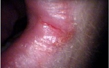
Mundwinkelentzündung
| Klassifikation nach ICD-10 | |
|---|---|
| K13.0 | Krankheiten der Lippen |
Eine Mundwinkelentzündung (auch: Mundwinkelrhagade, Cheilitis angularis, Angulus infectiosus oris, Perlèche [französisch pourlècher, sich die Lippen lecken][1] oder Faulecken) ist eine Erkrankung der Haut und Schleimhaut der Mundwinkel.[2][1][3]
Ätiologie
Es gibt derzeit (2016) kaum wissenschaftliche Studien über die Krankheitshäufigkeit (Prävalenz) und ihre Ursachen (Ätiologie).[1] Die Annahme, dass Mundwinkelentzündungen mit einer Vielzahl an lokalen und systemischen Faktoren zusammenhängen, beruht auf ärztlichen Erfahrungen.[1] Diese lokalen und systemischen Faktoren können einzeln oder kombiniert auftreten.
Lokale Ursachen
Im Vordergrund stehen, da am häufigsten zugrunde liegend, drei lokale Ursachen:[1]
- Lokale Kontaktdermatitis durch anatomisch bedingte mechanische oder chemische Reize
- Allergische Kontaktdermatitis
- Infektiöse Ursachen (zum Beispiel eine Pilzinfektion mit Candida albicans oder einer bakteriellen Infektion mit Staphylococcus aureus)[4]
Systemische Ursachen
Systemische Ursachen können andere Grunderkrankungen, Fehlernährung, Drogenmissbrauch oder Medikamente sein.[5] Es wird angenommen, dass bei etwa einem Viertel der Fälle die Ursache in einem Mangel an B-Vitaminen (Riboflavin (Vitamin B2), Pyridoxin (Vitamin B6), Biotin (Vitamin B7) und Cobalamin (Vitamin B12))[6] oder Eisen liegt.[5]
Es wird vermutet, dass Eisenmangel zu Immunschwäche führt und hierdurch opportunistische Candida-Infektionen auftreten.[5]
Ebenso ist Zinkmangel eine bekannte Ursache,[7] die außerdem zu Durchfall, Haarausfall und Dermatitis führt. Acrodermatitis enteropathica ist eine angeborene Stoffwechselstörung, die zu verminderter Zinkaufnahme führt und daher mit Mundwinkelentzündungen assoziiert ist.[5]
Mundwinkelentzündungen kommen am häufigsten in der dritten, fünften und sechsten Lebensdekade vor.[1] Sie haben bei Erwachsenen einen Anteil von 0,7 bis 3,8 % aller Schleimhautläsionen. Bei Kindern sind es 0,2 bis 15,1 % aller oraler Läsionen.[1]
Symptome
Symptome sind allgemeine Entzündungszeichen, Mazeration, Ulzeration und Verkrustung.[2] Eine Mundwinkelentzündung kann auf einer oder auf beiden Seiten gleichzeitig entstehen.[1] Der Beginn kann schleichend oder akut sein.[1] Der Verlauf kann spontan remittieren oder wiederkehrend sein.[1]
Therapie
Hauptpfeiler der ersten Behandlung ist es, die als lokalen Auslöser bekannten Ursachen für die Beeinträchtigung der Haut im Mundwinkelbereich zu entfernen.[1] Ziel ist es, einen chronischen Verlauf zu verhindern.[1]
Die Behandlung lokaler Ursachen umschließt:
- Sicherstellung des korrekten Sitzes und der korrekten Reinigung von Zahnersatz[1]
- Sicherstellung von korrekter Mundhygiene[1]
- Benutzung von Speichelersatzmitteln (Sialogoga), falls nötig[1]
- Schutzcreme als Barriere abends (z. B. Zinkoxid-Paste)[1]
Diese Maßnahmen reichen in der Regel aus, um eine Linderung zu erreichen.[1] Wenn alle Möglichkeiten, lokale Ursachen zu behandeln, erschöpft sind, sollten weniger häufige Ursachen einer Mundwinkelentzündung identifiziert werden.[5]
Siehe auch
Einzelnachweise
- ↑ a b c d e f g h i j k l m n o p q Kelly. K. Park, Robert T. Brodell, Stephen E. Helms: Angular cheilitis, part1: local etiologies. In: Cutis. Band 87, Nr. 6, Juni 2011, S. 289–295, PMID 21838086 (Review).
- ↑ a b Perlèche. (Nicht mehr online verfügbar.) In: Die Online Enzyklopädie der Dermatologie, Venerologie, Allergologie und Umweltmedizin. P. Altmeyer. Archiviert vom Original am 3. April 2015; abgerufen am 25. Juni 2016.
- ↑ Dorothea Terhorst (Hrsg.): Basics Dermatologie. 2. Auflage. Elsevier, Urban & Fischer, 2009, ISBN 978-3-437-42137-2, S. 129–130.
- ↑ W. C. Gonsalves, A. S. Wrightson, R. G. Henry: Common oral conditions in older persons. In: American family physician. Band 78, Nr. 7, Oktober 2008, S. 845–852, PMID 18841733 (Review).
- ↑ a b c d e Kelly K. Park, Robert T. Brodell, Stephen E. Helms: Angular cheilitis, part 2: nutritional, systemic, and drug-related causes and treatment. In: Cutis. Band 88, Nr. 1, Juli 2011, S. 27–32, PMID 21877503 (Review).
- ↑ Ulric Thomas Ruzicka (Hrsg.): Lehrbuch der Dermatologie und Venerologie. Ihr roter Faden durch Studium nach der neuen ÄAppO. Wissenschaftliche Verlagsgesellschaft, Stuttgart 2006, ISBN 3-8047-2178-8.
- ↑ D. Gaveau, F. Piette, A. Cortot, V. Dumur, H. Bergoend: [Cutaneous manifestations of zinc deficiency in ethylic cirrhosis]. In: Ann Dermatol Venereol. Band 114, Nr. 1, 1987, S. 39–53, PMID 3579131 (englisch).
Auf dieser Seite verwendete Medien
Autor/Urheber: Matthew Ferguson 57, Lizenz: CC BY-SA 3.0
Photograph of bilateral angular cheilitis in an elderly individual with false teeth, iron deficiency anemia and xerostomia
This patient signed a consent form a blank copy of which is pasted below:
Consent Form for Clinical Photography • Your clinician would like to take a photograph
• The image may be used for teaching purposes in a healthcare environment (e.g. a lecture or teaching session with students or junior doctors), in presentations for medical conferences, in professional medical publications (including journals and textbooks), or in websites (e.g. medical articles on Wikipedia under the Creative Commons Attribution-Share Alike 3.0 Unported copyright license*)
• The image will be taken on a mobile device and then promptly transferred to a secure hard drive along with an electronic copy of this form to be stored for reference
• You will NEVER be personally identifiable from the image, and you can view it after it has been taken to check you are happy with it
• Your personal information, such as name and address will NEVER be disclosed with the image. Very general details, such as your rough age and gender may be disclosed. Some medical details about what is shown in the image may also be disclosed, e.g. the diagnosis or appropriate terminology describing the appearance
I understand and consent to the above
Patient name ………………………………………… Signed (patient / parent / legal guardian) ………………………………………… Signed (clinician) ………………………………………… Date …………………………………………
CC BY-SA 3.0 for details please see:
https://creativecommons.org/licenses/by-sa/3.0/Autor/Urheber: Internet Archive Book Images, Lizenz: No restrictions
Identifier: diseasesofinfant01grif (find matches)
Title: The diseases of infants and children
Year: 1919 (1910s)
Authors: Griffith, J. P. Crozer (John Price Crozer), 1856-1941
Subjects: Infants Children Disease Pediatrics
Publisher: Philadelphia, London, W.B. Saunders company
Contributing Library: Columbia University Libraries
Digitizing Sponsor: Open Knowledge Commons
View Book Page: Book Viewer
About This Book: Catalog Entry
View All Images: All Images From Book
Click here to view book online to see this illustration in context in a browseable online version of this book.
Text Appearing Before Image:
^ studied by Lemaistre^ in1886 and called La Perleche, is not of very frequent occurrence. TheFrench name is derived from the tendency of the patient to lick the affectedareas (pour lecher). Etiology.—It effects especially early childhood, being rarely seen inadults; is favored by the presence of a state of debility and by unclean-liness; and would even appear to be contagious, since several members ofone family may be attacked, or it may occur epidemically in schools.A streptococcus has been described by Lemaistre.
Text Appearing After Image:
Fig. 226.—Erosions at the Angles of the Mouth.(Epstein, Jahrh. f. Kinderheilk., 1900, LI, 317.) Symptoms.—The lesions consist of small, fissure-like ulcers arrangedradially at the labial angle, generall)^ at both sides. The skin is red andswollen, and under the influence of the moistening by the constant applica-tion of the tongue a grayish ulcer of larger size may develop (Fig. 226).The lesions arc painful, but do not affect the child in other respects.The course is favorable and recovery takes place in from 2 to 4 weeksunder treatment, leaving no scars in the majority of instances; but with-out treatment the course may be prolonged and may become chronic.Except in severe cases there is no lymi)hatic involvement. Diagnosis.—^This is a matter of importance since the lesions ))ear aresemblance to the placjues of syphilis. Thej differ in that the fissuresnever extend into the mouth nor deeply into the tissues; are withoutinduration at the base; and are not strictly limited to the
Note About Images
Autor/Urheber: Lesion, Lizenz: CC BY-SA 3.0
Zoomed, Cropped and rm lable from original image: Photographic Comparison of a Canker Sore, Herpes, Angular Cheilitis and Chapped Lips..jpg


