Mumpsvirus
| Mumpsvirus | ||||||||||||||||||||
|---|---|---|---|---|---|---|---|---|---|---|---|---|---|---|---|---|---|---|---|---|
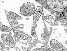
 Mumpsvirus in der TEM-Abbildung | ||||||||||||||||||||
| Systematik | ||||||||||||||||||||
| ||||||||||||||||||||
| Taxonomische Merkmale | ||||||||||||||||||||
| ||||||||||||||||||||
| Wissenschaftlicher Name | ||||||||||||||||||||
| Mumps orthorubulavirus | ||||||||||||||||||||
| Kurzbezeichnung | ||||||||||||||||||||
| MuV | ||||||||||||||||||||
| Links | ||||||||||||||||||||
|
Das Mumpsvirus (offiziell Mumps orthorubulavirus, manchmal auch als Paramyxovirus parotitis bezeichnet) ist ein nur beim Menschen vorkommendes Virus aus der Familie Paramyxoviridae und ist der Erreger des Mumps (Parotitis epidemica). Das Virus konnte erstmals 1945 von John Franklin Enders in bebrüteten Hühnereiern vermehrt und charakterisiert werden.[3] 1954 wurde die damals außergewöhnliche Morphologie des Mumpsvirus im Elektronenmikroskop erstmals untersucht.[4]
Das Mumpsvirus besitzt eine lipidhaltige Virushülle, die ein geknäuelt angeordnetes, helikales Kapsid umgibt. Diese Struktur erklärt die Empfindlichkeit des Mumpsvirus gegenüber milden Seifen und Austrocknung. Das Virus ist als einheitlicher Serotyp weltweit verbreitet und verfügt über kein tierisches Reservoir.
Morphologie

Das Virion des Mumpsvirus erscheint rund bis unregelmäßig mit einem mittleren Durchmesser von 150 nm. In der Virushülle befinden sich zwei Hüllproteine (F1 und F2), von denen das F1 eine Hämagglutinin- und Neuraminidase-Aktivität aufweist. F1 und F2 lagern sich als Heterodimer zusammen und bilden das so aktive Fusionsprotein zum Eintritt in die Zelle. Die Innenseite der Hülle ist von einem Matrixprotein bedeckt, das den Zusammenbau des Viruspartikels beim Austritt an der Zellmembran ermöglicht.
Das helikale Kapsid umschließt die virale RNA und besteht aus vier verschiedenen Kapsidproteinen, zum überwiegenden Teil dem N-Protein (N: Nukleokapsid). Die Anwesenheit des N-Proteins ist auch bei der Transkription der viralen RNA erforderlich. Im Inneren des Virions wird zusätzlich ein Molekül der viralen RNA-Polymerase verpackt, um unmittelbar nach Eintritt in die Zelle eine komplementäre (+)RNA zu synthetisieren.
Genom
Das Genom des Mumpsvirus ist ein einziger, linearer Strang RNA mit negativer Polarität (ss(-)RNA) und einer Länge von 15.384 nt. Das Genom beinhaltet neun Offene Leserahmen (ORF), die für acht virale Proteine codieren: Nukleo- (N), Phospho- (P), Matrix- (M), Fusionsprotein (F), großes Protein (L), V-Proteine sowie ein kleines, hydrophobes (SH) Protein.[5] Außerdem wird eine Hämagglutinin-Neuraminidase (HN) kodiert.[5] Das SH-Protein verhindert, dass die infizierte Zelle in die Apoptose geht. Das V-Protein ist nur in infizierten Zellen nachweisbar und verhindert die Aktivierung einer Interferon-induzierten antiviralen Antwort.
Die ORFs sind nacheinander ohne Überlappung angeordnet und werden von kurzen nichtcodierenden Bereichen getrennt. An den Enden des RNA-Stranges befinden sich weder eine 5'-Cap-Struktur noch ein Poly-A-Schwanz.
Subtypen und Impfstämme
Weltweit wurden mehrere genetisch leicht unterschiedliche Subtypen des Mumpsvirus isoliert, die sich jedoch weder in der Erkrankung noch in der serologischen Reaktion unterscheiden; das Mumpsvirus liegt damit trotz kleiner Varianten in nur einem weltweiten Serotyp vor. Einige natürliche oder in der Zellkultur gezüchtete Stämme werden in abgeschwächter Form als attenuierter Lebendimpfstoff zur Schutzimpfung verwendet (Mumpsimpfstoff). In Deutschland wird der Impfstoff überwiegend aus dem Stamm Jeryl-Lynn hergestellt, der in bebrüteten Hühnereiern vermehrt wird. Während die Mumpsinfektion eine lebenslange Immunität hinterlässt, vermag die einmalige Impfung eine solche nicht immer zu induzieren.[6] Die spezifischen Antikörper (IgG) gegen das Mumpsvirus sinken in ihrer Plasmakonzentration sehr rasch ab und sind im Verlaufe mehrere Jahre in den derzeitigen Testverfahren oft nur schlecht oder gar nicht mehr nachweisbar; dieses Verschwinden der Nachweisbarkeit des Mumps-IgG ist nicht unbedingt ein Zeichen nicht vorhandener zellulärer Immunität.[7]
- Spezies Mumpsvirus
- Subtyp Mumpsvirus Stamm Belfast
- Subtyp Mumpsvirus Stamm Bristol
- Subtyp Mumpsvirus Stamm Edinburg 2
- Subtyp Mumpsvirus Stamm Edinburg 4
- Subtyp Mumpsvirus Stamm Edinburg 6
- Subtyp Mumpsvirus Stamm Enders
- Subtyp Mumpsvirus Stamm Kilham
- Subtyp Mumpsvirus Stamm Matsuyama
- Subtyp Mumpsvirus Stamm RW
- Subtyp Mumpsvirus Stamm SBL
- Subtyp Mumpsvirus Stamm SBL-1
- Subtyp Mumpsvirus Stamm Takahashi
- Subtyp Mumpsvirus Stamm Jeryl-Lynn
- Subtyp Mumpsvirus Stamm L-Zagreb
- Subtyp Mumpsvirus Stamm Urabe
- Subtyp Mumpsvirus Stamm Miyahara-Vakzine
- Subtyp Mumpsvirus Stamm Urabe-Vakzine AM9
Meldepflicht
In Deutschland ist der direkte oder indirekte Nachweis vom Mumpsvirus namentlich meldepflichtig nach § 7 des Infektionsschutzgesetzes, soweit der Nachweis auf eine akute Infektion hinweist.
Quellen
- S. Mordrow, D. Falke, U. Truyen: Molekulare Virologie, Heidelberg Berlin, 2. Auflage 2003
- R. A. Lamb et al.: Genus Rubulavirus. In: C.M. Fauquet, M.A. Mayo et al.: Eighth Report of the International Committee on Taxonomy of Viruses, London, San Diego, 2005, S. 659f
Einzelnachweise
- ↑ a b ICTV: ICTV Taxonomy history: Akabane orthobunyavirus, EC 51, Berlin, Germany, July 2019; Email ratification March 2020 (MSL #35)
- ↑ ICTV Master Species List 2018b.v2. MSL #34, März 2019
- ↑ J. H. Levens und J. F. Enders: The hemoagglutinative properties of amniotic fluid from embryonated eggs infected with mumps virus. In: Science (New York, N.Y.). Band 102, Nr. 2640, 3. August 1945, S. 117–120, doi:10.1126/science.102.2640.117, PMID 17777358.
- ↑ B. G. Ray und R. H. Swain: An investigation of the mumps virus by electron microscopy. In: The Journal of Pathology and Bacteriology. Band 67, Nr. 1, Januar 1954, S. 247–252, doi:10.1002/path.1700670130, PMID 13152639.
- ↑ a b T. Betáková et al.: Overview of measles and mumps vaccine: origin, present, and future of vaccine production. In: Acta Virologica. Band 57, Nr. 2, 2013, S. 91–96, doi:10.4149/av_2013_02_91, PMID 23600866.
- ↑ Gaynor Watson-Creed et al.: Two successive outbreaks of mumps in Nova Scotia among vaccinated adolescents and young adults. In: CMAJ: Canadian Medical Association journal = journal de l'Association medicale canadienne. Band 175, Nr. 5, 29. August 2006, S. 483–488, doi:10.1503/cmaj.060660, PMID 16940266, PMC 1550754 (freier Volltext).
- ↑ Sari Jokinen et al.: Cellular immunity to mumps virus in young adults 21 years after measles-mumps-rubella vaccination. In: The Journal of Infectious Diseases. Band 196, Nr. 6, 15. September 2007, S. 861–867, doi:10.1086/521029, PMID 17703416.
Weblinks
Auf dieser Seite verwendete Medien
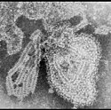
ID#: 8758 Description: This 1973 negative stained transmission electron micrograph (TEM) depicted the ultrastructural features displayed by the mumps virus. The mumps virus replicates in the upper respiratory tract and is spread through direct contact with respiratory secretions or saliva or through fomites, i.e., inanimate objects that are contaminated by the virus, and are subsequently handled. The infectious period or time that an infected person can transmit mumps to a non-infected person is from 3 days before symptoms appear to about 9 days after the symptoms appear. The incubation time, which is the period from when a person is exposed to virus to the onset of any symptoms, can vary from 16 to 18 days (range 12-25 days).
Content Providers(s): CDC/ Dr. F. A. Murphy Creation Date: 1973
Copyright Restrictions: None - This image is in the public domain and thus free of any copyright restrictions. As a matter of courtesy we request that the content provider be credited and notified in any public or private usage of this image.This 1977 thin sectioned transmission electron micrograph (TEM) depicted the ultrastructural details of the mumps virions that had been grown in a Vero cell culture.
Mumps is an acute viral illness caused by the mumps virus. Symptoms include fever, headache, muscle aches, tiredness, and loss of appetite, which are followed by swelling of salivary glands. The parotid salivary glands (which are located within your cheek, near your jaw line, below your ears) are most frequently affected.
Severe complications are rare. However, mumps can cause:
-inflammation of the brain and/or tissue covering the brain and spinal cord (encephalitis/meningitis)
-inflammation of the testicles (orchitis)
-inflammation of the ovaries and/or breasts (oophoritis and mastitis)
-spontaneous abortion
-deafness, usually permanentAus dem Virion freigesetzte Anteile des helikalen Kapsids.


