Methylomirabilis
| Methylomirabilis | ||||||||||||
|---|---|---|---|---|---|---|---|---|---|---|---|---|
 TEM-Aufnahme von gefriergetrockneten Methylomirabilis-Zellen im Dünnschnitt; Balken 200 nm.[1] | ||||||||||||
| Systematik | ||||||||||||
| ||||||||||||
| Wissenschaftlicher Name | ||||||||||||
| Methylomirabilis | ||||||||||||
| Ettwig et al., 2010 |
Candidatus Methylomirabilis[Anm. 1] ist eine Kandidatengattung gramnegativer Bakterien, in der Klade A des vorgeschlagenen Phylums NC10.[2][3][4] Dieses ist Mitglied oder Schwesterphylum der „Rokubacteria“.[5]
Als Ordnung der Gattung Methylomirabilis innerhalb des Phylums NC10 wurde der Name „Methylomirabilales“ vorgeschlagen,[6] als Familie Methylomirabilaceae.[2]
Typusgattung ist Ca. M. oxygeniiferaEttwig et al. 2010 syn. Ca. M. oxygeniifera corrig.Ettwig et al. 2010.[7]
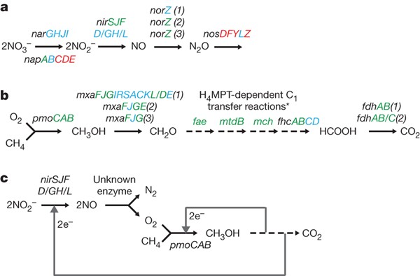
Diese Bakterien zeichnen sich durch ihre Fähigkeit aus, anaerobe Methanoxidation mit Nitritreduktion in anoxischen Umgebungen zu koppeln.[8][9] Um den Sauerstoff für die Methanoxidation zu erhalten, nutzt M. oxyfera einen intra-aeroben Weg per Reduktion von Nitrit (NO2) zu Distickstoff (N2) und Sauerstoff (O2).[10]

(a) Gelbe Pfeile zeigen auf Mikrokolonien.
(b) Die Schemazeichnung wurde auf der Grundlage des CLSM-Bildes links vorgeschlagen. Balken 20 μm.
Der Stamm mit dem vorgeschlagenen Speziesnamen Ca. M. sinica bildet Mikrokolonien. Die Bakterien selbst sind offenbar Stäbchen mit einem polygonalen Querschnitt, ebenso wie Ca. M. oxyfera.[11][1]
Etymologie
Der Name setzt sich zusammen aus dem Präfix „methylo“ (was Bezug nimmt auf die verstoffwechselte Methylgruppe) und der Endung „mirabilis“ (lateinisch für erstaunlich, seltsam).[3]
Anreicherung
Durch den Einsatz von elektronenmikroskopischer Techniken wurden angereicherte Zellen der Typusspezies Ca. M. oxyfera mit einer spezifischen polygonalen Zellform identifiziert. Im Gegensatz zu methanotrophen Proteobakterien weisen die M. oxyfera-Zellen keine (jedenfalls unter Laborbedingungen) keine Membranen innerhalb des Zellplasmas (intrazytoplasmatische Membranen) auf.[12] Die optimalen Wachstumsbereiche für Ca. M. oxyfera liegen zwischen pH 7–8 und 25–30 °C.[8] Die Zellhüllen von Ca. M. oxyfera sind Gram-negativ und haben einen Durchmesser von i. a. 0,25–0,5 μm bei einer Länge von 0,8–1,1 μm.[8][12]
Methan-Oxidation
Ca. M. oxyfera hat die Fähigkeit, Stickstoffoxid in Sauerstoff und Stickstoffgas zu disproportionieren. Dieser intermediäre Sauerstoff wird dann bei der Oxidation von Methan zu Kohlendioxid verwendet.[8][10]
- 3 CH4 + 8 NO2 + 8 H+ → 3 CO2 + 4 N2 + 10 H2O
Umweltbedeutung
Ca. M. oxyfera wurde in verschiedenen Umgebungen gefunden, darunter in den Böden von Reisfeldern in China,[13] in mehreren Fluss- und Seesedimenten,[14] und in Abwasserschlamm in den Niederlanden.[15]
Allgemein bewohnt Ca. M. oxyfera vermutlich Umgebungen mit hohen Stickstoff- und Methankonzentrationen im Übergangsbereich von oxischen und anoxischen Zonen. Man nimmt an, dass Ca. M. oxyfera und ähnliche Organismen signifikant zu den globalen Kohlenstoff- und Stickstoffkreisläufen beitragen. Diese Organismen könnten auch eine Rolle bei der Reduzierung der Nährstoffbelastung in Süßwasser-Ökosystemen spielen, die mit Düngemitteln kontaminiert wurden.[14]
Bakteriophagen
Bakteriophagen (Bakterienviren), die Ca. „Methylomirabilis“ (Ca. „M. oxyfera“ und eine weitere Spezies) infizieren, wurden 2016 von Lavinia Gambelli und Kollegen untersucht. Es wurden mehrere Phagen mit Kopf-Schwanz-Aufbau wie bei den Myoviren (Klasse Caudoviricetes) identifiziert, sowie ein schwanzloser Phage mit ikosaedrischem Kapsid. Bakteriophagen können auch in Bioreaktor-Anreicherungsanlagen schwerwiegende Auswirkungen auf die Bakterienpopulationen haben, was bei einer Anwendung der Mikroorganismen (z. B. in Abwasseraufbereitungsanlagen) berücksichtigt werden muss.[1]


Systematik
Zur Kandidatengattung Ca. „Methylomirabilis“ gehören nach der List of Prokaryotic names with Standing in Nomenclature (LPSN) und dem National Center for Biotechnology Information (NCBI) folgende Spezies:[3][4]
- „Ca. M. lanthanidiphila“Versantvoort et al. 2018, syn. „Ca. M. sp. lanth“[16]
- „Ca. M. Methylomirabilis limnetica“Graf et al. 2018, syn. „Ca. M. sp. Zug“[17] – im Zugersee, Schweiz
- „Ca. M. oxygeniifera“ corrig.Ettwig et al. 2010 Typus, syn. „Ca. M. oxyfera“Ettwig et al. 2010 (orthografische Variante), veraltet: „NC10 bacterium 'Dutch sediment'“

Nur beim NCBI sind gelistet:
- „Ca. M. sinica“He et al. 2016[11] – inklusive Stamm RS1
- „Ca. M. sp. BIN6“
- „Ca. M. sp. MW5-17“
- „Ca. M. sp. RS2“
- „Ca. M. sp. RS3“
Anmerkungen
- ↑ manchmal verschrieben als Methylomirabilish oder Methylomirabils (LPSN)
Einzelnachweise
- ↑ a b c d e f Lavinia Gambelli, Geert Cremers, Rob Mesman, Simon Guerrero, Bas E. Dutilh, Mike S. M. Jetten, Huub J. M. Op den Camp, Laura van Niftrik: Ultrastructure and Viral Metagenome of Bacteriophages from an Anaerobic Methane Oxidizing Methylomirabilis Bioreactor Enrichment Culture, in: Frontiers in Microbiology, Band 7, S. 1740, 8. November 2016, doi:10.3389/fmicb.2016.01740, ISSN 1664-302X
- ↑ a b Natalie June Gayner: River Bank Inducement Influence on a Shallow Groundwater Microbial Community and Its Effects on Aquifer Reactivity, Dissertation an der University of Wisconsin-Milwaukee (UWM), Dezember 2018. Insbes. APPENDIX D: W12 UNIQUE DNA AND RNA TAXA SPECIALISTS, Table 26: Unique W12 Specialists from DNA and RNA CLAM Results.
- ↑ a b c LPSN: Genus "Candidatus Methylomirabilis"
- ↑ a b NCBI: "Candidatus Methylomirabilis" Ettwig et al. 2010 (genus); graphisch: Candidatus Methylomirabilis, auf: Lifemap, NCBI Version.
- ↑ Natascha Menezes Bergo, Amanda Gonçalves Bendia, Juliana Correa Neiva Ferreira, Bramley J. Murton, Frederico Pereira Brandini, Vivian Helena Pellizari: Microbial Diversity of Deep-Sea Ferromanganese Crust Field in the Rio Grande Rise, Southwestern Atlantic Ocean, in: Environmental Microbiology, 16. Januar 2021, doi:10.1007/s00248-020-01670-y. Freier Preprint:
- Natascha Menezes Bergo, Amanda Gonçalves Bendia, Juliana Correa Neiva Ferreira, Bramley Murton, Frederico Pereira Brandini, Vivian Helena Pellizari: Microbial Diversity of Deep-Sea Ferromanganese Crust Field in the Rio Grande Rise, Southwestern Atlantic Ocean, auf: bioRxiv, 13. Juni 2020, doi:10.1101/2020.06.13.150011
- ↑ Léa Cabrol et al.: Anaerobic oxidation of methane and associated microbiome in anoxic water of Northwestern Siberian lakes, in: Science of The Total Environment, Band 736, 20. September 2020, 139588, doi:10.1016/j.scitotenv.2020.139588. Abschnitt 3.3
- ↑ LPSN: Species "Candidatus Methylomirabilis oxyfera" und Species "Candidatus Methylomirabilis oxygeniifera"
- ↑ a b c d Katharina F. Ettwig, Margaret K. Butler, Denis Le Paslier, Eric Pelletier, Sophie Mangenot, Marcel M. M. Kuypers, Frank Schreiber, Bas E. Dutilh, Johannes Zedelius, Dirk de Beer, Jolein Gloerich: Nitrite-driven anaerobic methane oxidation by oxygenic bacteria. In: Nature. 464. Jahrgang, Nr. 7288, März 2010, ISSN 1476-4687, S. 543–548, doi:10.1038/nature08883, PMID 20336137 (englisch, nature.com).
- ↑ Mohamed F. Haroon, Shihu Hu, Ying Shi, Michael Imelfort, Jurg Keller, Philip Hugenholtz, Zhiguo Yuan, Gene W. Tyson: Anaerobic oxidation of methane coupled to nitrate reduction in a novel archaeal lineage. In: Nature. 500. Jahrgang, Nr. 7464, August 2013, ISSN 1476-4687, S. 567–570, doi:10.1038/nature12375, PMID 23892779 (englisch, nature.com). Siehe insbes. Fig. 1.
- ↑ a b Ming L. Wu, Katharina F. Ettwig, Mike S. M. Jetten, Marc Strous, Jan T. Keltjens, Laura van Niftrik: A new intra-aerobic metabolism in the nitrite-dependent anaerobic methane-oxidizing bacterium Candidatus 'Methylomirabilis oxyfera'. In: Biochemical Society Transactions. 39. Jahrgang, Nr. 1, 1. Februar 2011, ISSN 0300-5127, S. 243–248, doi:10.1042/BST0390243, PMID 21265781 (englisch, portlandpress.com).
- ↑ a b Zhanfei He, Chaoyang Cai, Jiaqi Wang, Xinhua Xu, Ping Zheng, Mike S. M. Jetten, Baolan Hu: A novel denitrifying methanotroph of the NC10 phylum and its microcolony, in: Scientific Reports, Band 6, Nr. 32241, 1. September 2016, doi:10.1038/srep32241
- ↑ a b Ming L. Wu, Muriel C. F. van Teeseling, Marieke J. R. Willems, Elly G. van Donselaar, Andreas Klingl, Reinhard Rachel, Willie J. C. Geerts, Mike S. M. Jetten, Marc Strous, Laura van Niftrik: Ultrastructure of the Denitrifying Methanotroph "Candidatus Methylomirabilis oxyfera," a Novel Polygon-Shaped Bacterium. In: Journal of Bacteriology. 194. Jahrgang, Nr. 2, 15. Januar 2012, ISSN 0021-9193, S. 284–291, doi:10.1128/JB.05816-11, PMID 22020652, PMC 3256638 (freier Volltext) – (englisch).
- ↑ Zhanfei He, Chaoyang Cai, Jiaqi Wang, Xinhua Xu, Ping Zheng, Mike S. M. Jetten, Baolan Hu: A novel denitrifying methanotroph of the NC10 phylum and its microcolony. In: Scientific Reports. 6. Jahrgang, Nr. 1, Oktober 2016, ISSN 2045-2322, S. 32241, doi:10.1038/srep32241, PMID 27582299, PMC 5007514 (freier Volltext) – (englisch).
- ↑ a b Li-Dong Shen, Zhan-Fei He, Qun Zhu, Dong-Qing Chen, Li-Ping Lou, Xiang-Yang Xu, Ping Zheng, Bao-Lan Hu: Microbiology, ecology, and application of the nitrite-dependent anaerobic methane oxidation process. In: Frontiers in Microbiology. 3. Jahrgang, 2012, ISSN 1664-302X, S. 269, doi:10.3389/fmicb.2012.00269, PMID 22905032, PMC 3408237 (freier Volltext) – (frontiersin.org).
- ↑ Francisca A. Luesken, Theo A. van Alen, Erwin van der Biezen, Carla Frijters, Ger Toonen, Christel Kampman, Tim L. G. Hendrickx, Grietje Zeeman, Hardy Temmink, Marc Strous, Huub J. M. Op den Camp: Diversity and enrichment of nitrite-dependent anaerobic methane oxidizing bacteria from wastewater sludge. In: Applied Microbiology and Biotechnology. 92. Jahrgang, Nr. 4, November 2011, ISSN 0175-7598, S. 845–854, doi:10.1007/s00253-011-3361-9, PMID 21667086, PMC 3198195 (freier Volltext) – (englisch).
- ↑ Wouter Versantvoort, Simon Guerrero-Cruz, Daan R. Speth, Jeroen Frank, Lavinia Gambelli, Geert Cremers, Theo van Alen, Mike S. M. Jetten, Boran Kartal, Huub J. M. Op den Camp, Joachim Reimann: Comparative Genomics of Candidatus Methylomirabilis Species and Description of Ca. Methylomirabilis Lanthanidiphila. In: Frontiers in Microbiology. 9. Jahrgang, 2018, ISSN 1664-302X, S. 1672, doi:10.3389/fmicb.2018.01672, PMID 30140258, PMC 6094997 (freier Volltext) – (englisch).
- ↑ Jon S. Graf, Magdalena J. Mayr, Hannah K. Marchant, Daniela Tienken, Philipp F. Hach, Andreas Brand, Carsten J. Schubert, Marcel M. M. Kuypers, Jana Milucka: Bloom of a denitrifying methanotroph, ‘Candidatus Methylomirabilis limnetica’, in a deep stratified lake, in: sfam environmental biology, Band 20, Nr. 7, Juli 2018, S. 2598–2614, doi:10.1111/1462-2920.14285
Auf dieser Seite verwendete Medien
Autor/Urheber: Lavinia Gambelli, Geert Cremers, Rob Mesman, Simon Guerrero, Bas E. Dutilh, Mike S. M. Jetten, Huub J. M. Op den Camp, Laura van Niftrik, Lizenz: CC BY-SA 4.0
Transmission electron micrographs of high-pressure frozen, freeze-substituted, resin-embedded, and thin-sectioned Methylomirabilis cells taken from a bioreactor enrichment culture. (A,B) Infected Methylomirabilis cells (white arrow in A) are in clusters among non-infected cells (black arrows in A). The bacteriophage (white arrow in B) has a hexagonal shape and an internal electron dense core. (C,D) The bacteriophages are organized in a highly packed formation (white arrow in D). The replication and assembly of bacteriophages causes the Methylomirabilis cell to swell and eventually the cell wall breaks (black arrows in D). (E,F) Lysed Methylomirabilis cell releasing the viral progeny. (B,D,F) are enlargements of (A,C,E), respectively. Scale bars; 200 nm.
Autor/Urheber: Zhanfei He, Chaoyang Cai, Jiaqi Wang, Xinhua Xu, Ping Zheng, Mike S. M. Jetten, Baolan Hu, Lizenz: CC BY-SA 4.0
Confocal laser scanning microscope (CLSM) images of the microcolonies of NC10 bacteria.
Several techniques were used to visualize NC10 bacteria anf then merged together (overlay with false color). NC10 bacteria appear in yellow in the merged images. The dense matter surrounding the microcolonies is indicated by white arrows. Bar = 5 μm.
‘Candidatus Methylomirabilis sinica’ (M. sinica) is the proposed name of this species.
Autor/Urheber: Lavinia Gambelli, Geert Cremers, Rob Mesman, Simon Guerrero, Bas E. Dutilh, Mike S. M. Jetten, Huub J. M. Op den Camp, Laura van Niftrik, Lizenz: CC BY-SA 4.0
Snapshots of electron tomograms and models of free and intracellular bacteriophages infecting Methylomirabilis cells. Tomogram (A) and model (B) of an infected Methylomirabilis cell. Most bacteriophages have the capsid (blue) assembled around the electron dense core (green). Some bacteriophages are still in the process of assembly and only consist of the electron dense core (pink). The cell is swollen and the cytoplasmic membrane (dark blue) is broken at many places (arrows). The cell wall (yellow) is still intact. All green electron dense cores were surrounded by a capsid, but not all capsids were modeled for reasons of clarity. Tomogram (C) and isosurface density model (D) of two free bacteriophages showing the icosahedral capsid (blue) and electron dense core (yellow). Tomogram (E) and Chimera model (F) showing two free bacteriophages. The electron dense core is enclosed by a putative membrane (arrows).
Autor/Urheber: Zhanfei He, Chaoyang Cai, Jiaqi Wang, Xinhua Xu, Ping Zheng, Mike S. M. Jetten, Baolan Hu, Lizenz: CC BY-SA 4.0
A confocal laser scanning microscope (CLSM) image (a) and a floc conceptual model (b) of NC10 bacteria and other bacteria in the culture. NC10 bacteria appear in yellow (a) for the double hybridization of NC10 specific primer S-*-DBACT-1027-a-A-18 (red) and EUB I-III mix (green) (microcolonies are indicated by yellow arrows); and other bacteria are in green (a) for single hybridization of EUB I-III mix (two cells of them are indicated by green arrows). The floc conceptual model was proposed based on the CLSM image left. Scale bar is 20 μm.
‘Candidatus Methylomirabilis sinica’ (M. sinica) is the proposed name of the NC10 species.
Autor/Urheber: Lavinia Gambelli, Geert Cremers, Rob Mesman, Simon Guerrero, Bas E. Dutilh, Mike S. M. Jetten, Huub J. M. Op den Camp, Laura van Niftrik, Lizenz: CC BY-SA 4.0
Transmission electron micrographs of high-pressure frozen and freeze-etched Methylomirabilis cells taken from a bioreactor enrichment culture. (A) Cross-section of a non-infected (left) and infected (right) Methylomirabilis cell. (B) The bacteriophages (white arrows) contain a proteinaceous capsid. The capsid is made of triangular faces built by capsomeres. Concave (dashed black arrow) and convex (black arrow) printing of the internal core is visible in two of the viral particles. Scale bars; 200 nm.
Autor/Urheber: Lavinia Gambelli, Geert Cremers, Rob Mesman, Simon Guerrero, Bas E. Dutilh, Mike S. M. Jetten, Huub J. M. Op den Camp, Laura van Niftrik. Modification: combination with iset from same image, scale bar moved, color changed to black., Lizenz: CC BY-SA 4.0
Transmission electron micrographs of high-pressure frozen, freeze-substituted, resin-embedded, and thin-sectioned Methylomirabilis cells taken from a bioreactor enrichment culture. This image (and inset) shows non-infected cells (black arrows). Scale bar; 200 nm.