Kinetoplastea
| Kinetoplastea | ||||||||||||
|---|---|---|---|---|---|---|---|---|---|---|---|---|
 | ||||||||||||
| Systematik | ||||||||||||
| ||||||||||||
| Wissenschaftlicher Name | ||||||||||||
| Kinetoplastea | ||||||||||||
| Honigberg, 1963 |
Die Kinetoplastea (Syn.: Kinetoplastida) bilden eine Klasse geißeltragender einzelliger Organismen (Flagellaten); sie gehören zu den Euglenozoa. Viele Kinetoplasten sind Parasiten.
Beschreibung
Die Kinetoplastea haben ein einziges großes Mitochondrium, in dem sich der sogenannte Kinetoplast befindet: eine große Menge an fibrillärer DNA (kDNA), der oft in enger Beziehung zur Basis der Geißel steht.
Ökologie
Viele Kinetoplastea sind parasitär, wie zum Beispiel die Art Trypanosoma brucei, ein Parasit im Menschen, der die Schlafkrankheit auslöst. Überträger ist hier die Tsetse-Fliege. Eine andere, dem Menschen gefährliche, Kinetoplastideaart ist Trypanosoma cruzi, die die Chagas-Krankheit auslöst (Siehe auch: Trypanosomen).
Systematik


Traditionell wurden die Kinetoplastea in die beiden Unterordnungen Bodonida und Trypanosomida unterteilt. Adl et al. unterteilen aufgrund neuer Erkenntnisse die Gruppe folgendermaßen (aufgeführt ist jeweils eine Auswahl an Gattungen):
- Prokinetoplastina
- Apiculatamorpha (Gattung incertae sedis)
- Ichthyobodonidae (mit Perkinsellia und Ichthyobodo, dazu I. necator)
- Metakinetoplastina
- Neobodonida (mit Dimastigella, Neobodo, Rhynchobodo und Rhynchomonas)
- Parabodonida (mit Parabodo und Trypanoplasma)
- Eubodonida (mit Bodo)
- Trypanosomatida (mit Leishmania und Trypanosoma)
Literatur
- Sina M. Adl und 27 weitere Autoren: The New Higher Level Classification of Eukaryotes with Emphasis on the Taxonomy of Protists. The Journal of Eukaryotic Microbiology, Band 52, 2005, S. 399–451. doi:10.1111/j.1550-7408.2005.00053.x.
- Dmitri A Maslov, Sergei A Podlipaev und Julius Lukeš: Phylogeny of the Kinetoplastida: Taxonomic Problems and Insights into the Evolution of Parasitism. Memórias do Instituto Oswaldo Cruz, Band 6, 2001, S. 397–402.
Weblinks
Auf dieser Seite verwendete Medien
Autor/Urheber: P. M. Sachertt Mendes F. M. Lansac-Tôha B. R. Meira F. R. Oliveira L. F. M. Velho F. A. Lansac-Tôha, Lizenz: CC BY-SA 4.0
Neobodo designis (Skuja, 1948) Moreira et al., 2004. (A) Schematic drawing; (B) Photo, where the arrow indicates the apical flagellar pocket. Scale of 5 µm.
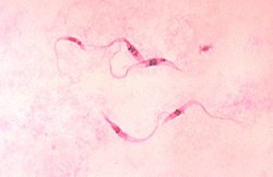
Nachweis von Trypanosoma cruzi.
Autor/Urheber: P. M. Sachertt Mendes F. M. Lansac-Tôha B. R. Meira F. R. Oliveira L. F. M. Velho F. A. Lansac-Tôha, Lizenz: CC BY-SA 4.0
Rhynchobodo simius Patterson & Simpson, 1996. (A) Schematic drawing; (B) Photo. Scale of 5 µm.
Autor/Urheber: P. M. Sachertt Mendes F. M. Lansac-Tôha B. R. Meira F. R. Oliveira L. F. M. Velho F. A. Lansac-Tôha, Lizenz: CC BY-SA 4.0
Dimastigella trypaniformis Sandon, 1928. (A) and (B) Schematic drawing; (C) and (D) Photos, where the arrow indicates the flagella adjacent to the cell. Scale of 5 µm.


