Heterocapsa
| Heterocapsa | ||||||||||||
|---|---|---|---|---|---|---|---|---|---|---|---|---|
 REM-Aufnahme zweier Zellen von H. rotunda vom Skagerrak (Schwedische Küste) | ||||||||||||
| Systematik | ||||||||||||
| ||||||||||||
| Wissenschaftlicher Name | ||||||||||||
| Heterocapsa | ||||||||||||
| Stein, 1883 |
Heterocapsa ist eine Gattung von Dinoflagellaten in der Familie Heterocapsaceae[2] (früher zu Pteridinidae).[3] Die Erstbeschreibung der Gattung stammt von F. Stein aus dem Jahr 1883.[3]
Heterocapsa ist Bestandteil des Meeresplanktons.[4] Die Gattung ist kosmopolitisch (weltweit verbreitet), hauptsächlich in Küstengewässern.[2][4]
Beschreibung
Die Dinoflagellaten der Gattung Heterocapsa sind mit 20–40 µm vergleichsweise mittelgroß. Die Einzeller sind biflagellat (doppelt begeißelt) und thecat (d. h. sie haben eine feste Hülle). Ihre Form ist unregelmäßig spindelförmig oder eiförmig mit einem mittelgroßen und runden Cingulum (Querfurche, auch „Gürtel“ genannt); der Sulcus (Längsfurche) ist auf das Hypokon beschränkt. Das genaue Muster der Thecaplatten ist schwer zu bestimmen und umstritten. Die von Balech (1988) veröffentlichte Formel lautet: Po, 4’, 2a, 7”, 6C, 5‴, 2⁗ und 4S.[5] Eine neuere Analyse bei H. minima findet sich bei Lee et al. (2019).[6] Wie bei der Gattung Cachonina (in der Schwesterfamilie Peridiniaceae)[7] befinden sich die Körperschuppen in einer einzigen Schicht auf der Zelloberfläche außerhalb der Thecalplatten. Die Individuen besitzen außer einem eiförmigen Zellkern ggf. zahlreiche Chloroplasten,[4] oder auch nur einen einzigen.[6]
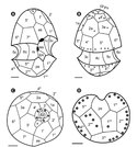
![Linkes Bild: REM-Aufnahmen von vegetativen Zellen von H. minima HMMJ1604:[6] (A) Ventrale Ansicht mit Epitheka, Cingulum (c), Sulcus (as, las, rs, lps und ps) und Hypotheka. (B) Ventral-linke Seitenansicht mit Epitheka, Cingulum (c), Sulcus (as, las, lps und ps) und Hypotheka. (C) Dorsale Ansicht mit Epitheka, Cingulum (c) und Hypotheka. (D) Ventral-rechtsseitige Ansicht mit Epitheka, Cingulum (c), Sulcus (as, rs, lps und ps) und Hypotheka. (E) Vergrößerung von (A) mit Sulcus (as, las, rs, lps und ps) (F) Apikaler Porenkomplex (Po, cp, ?, x) und in der Po-Platte angeordnete Thecalporen.](http://upload.wikimedia.org/wikipedia/commons/thumb/1/13/Algae-2019-34-1-7f3.tif/lossy-page1-260px-Algae-2019-34-1-7f3.tif.jpg)
(A) Ventrale Ansicht mit Epitheka, Cingulum (c), Sulcus (as, las, rs, lps und ps) und Hypotheka.
(B) Ventral-linke Seitenansicht mit Epitheka, Cingulum (c), Sulcus (as, las, lps und ps) und Hypotheka.
(C) Dorsale Ansicht mit Epitheka, Cingulum (c) und Hypotheka.
(D) Ventral-rechtsseitige Ansicht mit Epitheka, Cingulum (c), Sulcus (as, rs, lps und ps) und Hypotheka.
(E) Vergrößerung von (A) mit Sulcus (as, las, rs, lps und ps)
(F) Apikaler Porenkomplex (Po, cp, ?, x) und in der Po-Platte angeordnete Thecalporen.
![(G) Apikale Ansicht mit dem apikalen Porenkomplex (Po, cp, ?, x), Epitheka und Sulcus (as). (H) Antapikale Ansicht mit Hypotheka und Sulcus (ps). as, anteriorer Sulcus; las, linker anteriorer Sulcus; rs, rechter Sulcus; lps, linker posteriorer Sulcus; ps, posteriorer Sulcus; Po, apikale Porenplatte; cp, Deckplatte; x, Kanalplatte; ?, die zusätzliche Struktur, die als Scharnier/Verbindung dient. Rechtes Bild: Zeichnung einiger Ansichten von links:[6] (A) wie (A) links, (B) wie (C) links, (C) wie (G) links, (D) wie (H) links, –––––––– Balken jeweils 1 μm.](http://upload.wikimedia.org/wikipedia/commons/thumb/5/53/Algae-2019-34-1-7f4.tif/lossy-page1-323px-Algae-2019-34-1-7f4.tif.jpg)
(H) Antapikale Ansicht mit Hypotheka und Sulcus (ps). as, anteriorer Sulcus; las, linker anteriorer Sulcus; rs, rechter Sulcus; lps, linker posteriorer Sulcus; ps, posteriorer Sulcus; Po, apikale Porenplatte; cp, Deckplatte; x, Kanalplatte; ?, die zusätzliche Struktur, die als Scharnier/Verbindung dient.
Rechtes Bild: Zeichnung einiger Ansichten von links:[6]
(A) wie (A) links,
(B) wie (C) links,
(C) wie (G) links,
(D) wie (H) links,
––––––––
Balken jeweils 1 μm.
Vermehrung
Die Zellteilung findet im Geißelstadium statt, wobei sich die Tochterzellen die Theca der Mutterzelle teilen; geschlechtliche Fortpflanzung wurde nicht beobachtet.[4]
Arten


A: BF-Aufnahme einer lebenden Zelle
B, C: REM-Aufnahmen. Pfeile: Schuppen.
Mats Kuylenstierna[1]

Janina Kownacka[1]

Mats Kuylenstierna[1]
Die Systematik der Gattung Heterocapsa ist wie folgt:[4][2][8][1][9]
Familie Heterocapsaceae R.A. Fensome, F.J.R. Taylor, G. Norris, W.A.S. Sarjeant, D.I. Wharton & G.L. Williams
- Gattung Heterocapsa Stein, 1883 (gleich mit Heterocapsa J. Massart, 1920); Spezies:
- H. arctica T. Horiguchi, 1997(AGNWµ)
- Subspezies H. arctica subsp. frigida Rintala & G. Hällfors, 2010(NWµ)
- H. bohaiensis J. Xiao & Y. Li, 2018(AW)
- H. busanensis H. Choi & S. Kim(NA)
- H. chattonii (Biecheler) P. H. Campbell, 1973(AW)
- H. circularisquama Horiguchi, 1995(ANW)
- H. claromecoensis Sunesen, Rodríguez, Tillmann & Sar(AN)
- H. horiguchii Iwataki, Takayama & Matsuoka, 2002(AW)
- H. huensis M. Iwataki & Matsuoka(AN)
- H. illdefina (Herman & Sweeney) L. C. Morrill & Loeblich III, 1981(ANW)
- H. kollmeriana M. J. Swift & McLaughlin, 1970(AW)
- H. lanceolata Iwataki & Fukuyo, 2002(ANW)
- H. minima Pomroy, 1989(ANGWµ)
- H. niei (A. R. Loeblich III) L. C. Morrill & Loeblich III, 1981(AGNWµ)
- H. orientalis Iwataki, Botes & Fukuyo, 2003(ANW)
- H. ovata Iwataki & Fukuyo, 2003(AW)
- H. pacifica Kofoid, 1907(AW)
- H. psammophila M. Tamura, M. Iwataki & M. Horiguchi, 2006(ANW)
- H. pseudotriquetra Iwataki, G. Hansen & Fukuyo, 2002(ANW)
- H. pygmaea A. R. Loebl. et al., 1981(ANGW)
- H. rotundata (Lohmann) Hansen, 1995(AGNWµ)
- H. steinii Tillmann, Gottschling, Hoppenrath, Kusber & Elbrächter, 2017(ANW) – Typus (neu)[10]
- H. umbilicata Stein, 1883(AW)
Verschiebungen:
- H. quadridentata F. Stein, 1883; zu Peridinium quadridentatum (F. Stein) Gert Hansen, 1995
- H. triquetra (Ehrenberg) F. Stein, 1883(GNµ) (früher Glenodinium triquetrum Ehrenberg, Peridinium triquetra (Ehrenb.) Lebour) – Typus (alt); zu Kryptoperidinium triquetrum (Ehrenberg) U.Tillmann, M. Gottschling, M.Elbrächter, W.-H.Kusber & M.Hoppenrath, 2019(AW)[10]
Anmerkungen:
Algenblüten
H. triquetra (jetzt Kryptoperidinium triquetrum) kann dichte Algenblüten mit mehreren Millionen Zellen pro Liter bilden, die in Küstengewässern zu Verfärbungen führen. Eventuelle Fischsterben im Zusammenhang mit Heterocapsa-Blüten werden eher auf Sauerstoffmangel durch Zersetzung abgestorbener Dinoflagellaten als auf Algentoxine zurückgeführt.[4]
Viren
.jpg)
H. circularisquama wird parasitiert von der Virusspezies Heterocapsa circularisquama DNA virus 01 (HcDNAV),[12] einzige Art in der Gattung Dinodnavirus Spezies und Gattung dieser Doppelstrang-DNA-Viren sind zwar vom International Committee on Taxonomy of Viruses (ICTV) bestätigt, aber bisher (Stand Juli 2021) keinen höheren Taxa zugewiesen.[13]
Die Virusspezies wurde ursprünglich in der Familie Phycodnaviridae (Ordnung Algavirales) von Riesenviren der Klasse Megaviricetes (Phylum Nucleocytoviricota/NCLDV) vermutet. Untersuchungen des Genoms der Dinodnaviren haben aber gezeigt, dass die Gattung eher zur Familie Asfarviridae (heute in der Ordnung Asfuvirales, Klasse Pokkesviricetes) der NCLDV-Riesenviren gehört,[14] ähnlich wie die vorgeschlagenen Taxa „Faustovirus“, „Pacmanvirus“ und „Kaumoebavirus“. Näheres siehe „Faustovirus“ §Äußere Systematik.
Ein weiteres Virus dieser Algenspezies ist Heterocapsa circularisquama RNA virus 01 (HcRNAV01). Dies ist ein Einzelstrang-RNA-Virus positiver Polarität (Gattung Dinornavirus) in der Ordnung Sobelivirales der Orthornavirae.[15][12]
Einzelnachweise
- ↑ a b c d e f Nordic Microalgae: Heterocapsa Stein, auf: Nordic Microalgae and aquatic protozoa, Swedish Meteorological and Hydrological Institute (SMHI)
- ↑ a b c d Heterocapsa Stein, 1883. In: www.gbif.org. Abgerufen am 25. April 2021 (englisch).
Fotos von H. triquetra, jetzt Kryptoperidinium triquetrum - ↑ a b Friedrich Ritter von Stein: Die Naturgeschichte der arthrodelen Flagellaten. In: Der Organismus der Infusionsthiere. III. Abteilung: der arthrodelen Flagellaten oder Geisselinfusorien, II. Hälfte. Wilhelm Engelmann, Leipzig 1859, S. 1–30, insbesondere S. 13 (Textarchiv – Internet Archive).
- ↑ a b c d e f g AlgaeBase: Heterocapsa F.Stein, 1883
- ↑ Enrique Balech: Los dinoflagelados del Atlántico sudoccidental, Publicationes Especiales, Instituto Español de Oceanografia, Ministerio de Agricultura Pesca y Alimentation, Madrid, Nr. 1, 1988, PDF (spanisch). Hier: S. 158
- ↑ a b c d e Sung Yeon Lee, Hae Jin Jeong, Ji Eun Kwon, Ji Hyun You, So Jin Kim, Jin Hee Ok, Hee Chang Kang, Jae Yeon Park: First report of the photosynthetic dinoflagellate Heterocapsa minima in the Pacific Ocean: morphological and genetic characterizations and the nationwide distribution in Korea. In: Algae. Band 34, Nr. 1, 2019, ISSN 1226-2617, S. 7–21, doi:10.4490/algae.2019.34.2.28.
- ↑ AlgaeBase: Cachonina A.R.Loeblich III, 1968
- ↑ a b NCBI: Heterocapsa Stein, 1883 (genus); graphisch: Heterocapsa, auf: Lifemap, NCBI Version.
- ↑ a b WoRMS: Heterocapsa Stein, 1883
- ↑ a b Marc Gottschling, Urban Tillmann, Malte Elbrächter, Wolf-Henning Kusber, Mona Hopenrath: Glenodinium triquetrum Ehrenb. is a species not of Heterocapsa F.Stein but of Kryptoperidinium Er.Lindem. (Kryptoperidiniaceae, Peridiniales). In: Phytotaxa. Band 391, Nr. 2, 1. Februar 2019, ISSN 1179-3163, S. 155–158, doi:10.11646/phytotaxa.391.2.11.
- ↑ NCBI: unclassified Heterocapsa (list)
- ↑ a b Mohammadreza Sadeghi, Yuji Tomaru, Tero Ahola: RNA Viruses in Aquatic Unicellular Eukaryotes. In: Viruses. Band 13, Nr. 3, 2021, S. 362, doi:10.3390/v13030362, PMID 33668994 (Special Issue Viruses of Microbes 2020: The Latest Conquests on Viruses of Microbes).
- ↑ ICTV: ICTV Taxonomy history: Heterocapsa circularisquama DNA virus 01
- ↑ Hiroyuki Ogata, Kensuke Toyoda, Yuji Tomaru, Natsuko Nakayama, Yoko Shirai, Jean-Michel Claverie, Keizo Nagasaki: Remarkable sequence similarity between the dinoflagellate-infecting marine girus and the terrestrial pathogen African swine fever virus. In: Virology Journal. Band 6, Nr. 1, 2009, ISSN 1743-422X, S. 178, doi:10.1186/1743-422X-6-178, PMID 19860921.
- ↑ ICTV: ICTV Taxonomy history: Heterocapsa circularisquama RNA virus 01
Auf dieser Seite verwendete Medien
Autor/Urheber: Photographer: Janina Kownacka (National Marine Fisheries Research Institute, HELCOM-PEG) via Sveriges meteorologiska och hydrologiska institut (SMHI), Lizenz: CC BY-SA 3.0
Heterocapsa rotundata (Lohmann) Hansen, 1995 from Baltic Sea - The Gulf of Gdansk (autumn 2007). Light microscopy, Lugols iodine.

Autor/Urheber: Sung Yeon Lee, Hae Jin Jeong, Ji Eun Kwon, Ji Hyun You, So Jin Kim, Jin Hee Ok, Hee Chang Kang, Jae Yeon Park, Lizenz: CC BY-SA 3.0
Micrographs of living cells in the clonal culture of Heterocapsa minima HMMJ1604, taken by light microscopy. (A) H. minima cell showing the conical epitheca and rounded hypotheca with a deeply excavated cingulum. (B) A pyrenoid with starch sheath (asterisk) is positioned in the epitheca, while the large ellipsoid nucleus (N) is positioned in the central part of cell. The chloroplast lobe of a single chloroplast is positioned near the surface of a cell (arrow). (C) A large nucleus (N) and a small red accumulation body (arrowhead). Scale bars represent: A–C, 5 μm.
Autor/Urheber: Yoshihito Takano, Yuji Tomaru, Keizo Nagasaki / Viruses 2018. doi:10.3390/v10100554. This is an open access article distributed under the Creative Commons Attribution License, Lizenz: CC BY-SA 4.0
Figure 1 ABCDEHIJ. FE-SEM images of HcDNAV virions and virus-infected Heterocapsa circularisquama cells.
Autor/Urheber: Photographer: Mats Kuylenstierna, Maria Kuylenstierna, Bengt Karlson (University of Gothenburg) via Sveriges meteorologiska och hydrologiska institut (SMHI), Lizenz: CC BY-SA 3.0
Heterocapsa rotundata (Lohmann) Hansen, 1995 from Skagerrak-Kattegat. A living cell (BF); arrowhead: pyrenoid
Autor/Urheber: Photographer: Mats Kuylenstierna, Maria Kuylenstierna, Bengt Karlson (Dept. of Marine botany, University of Gothenburg) via Sveriges meteorologiska och hydrologiska institut (SMHI), Lizenz: CC BY-SA 3.0
Heterocapsa rotundata (Lohmann) Hansen, 1995 from Skagerrak - Swedish coast. SEM image of two cells. Notice the very thin plates. arrowhead: scale. Stain: Sputter coated
Autor/Urheber: Sung Yeon Lee, Hae Jin Jeong, Ji Eun Kwon, Ji Hyun You, So Jin Kim, Jin Hee Ok, Hee Chang Kang, Jae Yeon Park, Lizenz: CC BY-SA 3.0
Drawings of vegetative cells of Heterocapsa minima HMMJ1604, showing the external morphology. (A) Ventral view. (B) Dorsal view. (C) Apical view. (D) Antapical view. Scale bars represent: A–D, 1 μm. as, anterior sulcal; las, left anterior sulcal; rs, right sulcal; lps, left posterior sulcal; ps, posterior sulcal; Po, apical pore plate; cp, cover plate; x, canal plate; ?, the extra structure acting as a hinge or connection.

Autor/Urheber: Sung Yeon Lee, Hae Jin Jeong, Ji Eun Kwon, Ji Hyun You, So Jin Kim, Jin Hee Ok, Hee Chang Kang, Jae Yeon Park, Lizenz: CC BY-SA 3.0
REM-Aufnahmen von vegetativen Zellen von Heterocapsa minima HMMJ1604.
(A) Ventrale Ansicht mit Epitheka, Cingulum (c), Sulcus (as, las, rs, lps und ps) und Hypotheka.
(B) Ventral-linke Seitenansicht mit Epitheka, Cingulum (c), Sulcus (as, las, lps und ps) und Hypotheka.
(C) Dorsale Ansicht mit Darstellung der Epitheka, des Cingulums (c) und der Hypotheka.
(D) Ventral-rechtsseitige Ansicht mit Epitheka, Cingulum (c), Sulcus (as, rs, lps und ps) und Hypotheka.
(E) Vergrößerung von (A) mit detaillierter Ansicht des Sulcus (as, las, rs, lps und ps).
(F) Apikaler Porenkomplex (Po, cp, ?, x) und in der Po-Platte angeordnete Thekalporen.
(G) Apikale Ansicht mit dem apikalen Porenkomplex (Po, cp, ?, x), Epitheka und Sulcus (as).
(H) Antapikale Ansicht mit Hypotheka und Sulcus (ps). as, anteriorer Sulcus; las, linker anteriorer Sulcus; rs, rechter Sulcus; lps, linker posteriorer Sulcus; ps, posteriorer Sulcus; Po, apikale Porenplatte; cp, Deckplatte; x, Kanalplatte; ?, die zusätzliche Struktur, die als Scharnier oder Verbindung dient.
Die Skalenbalken zeigen jeweils 1 μm.
Autor/Urheber: Photographer: Mats Kuylenstierna, Maria Kuylenstierna (University of Gothenburg) via Sveriges meteorologiska och hydrologiska institut (SMHI), Lizenz: CC BY-SA 3.0
Heterocapsa minima Pomroy, 1989 from Skagerrak - Swedish coast. A : A living cell (BF - Bright-field); B, C: Cells (SEM), arrowheads: scales. Preservation: Glutardialdehyde