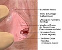
Glandula vestibularis major

Die Glandula vestibularis major (Mehrzahl Glandulae vestibulares majores; lateinisch für „große [Scheiden-]Vorhofdrüse“, auch Bartholin-Drüse oder Bartholin[i]sche Drüse) ist eine paarige akzessorische Geschlechtsdrüse der Frau. Sie mündet in den Scheidenvorhof zwischen den kleinen Schamlippen (Labia minora). Die Bezeichnung Bartholin-Drüse geht auf den dänischen Anatomen Caspar Bartholin d. J. (1655–1738) zurück.[1] Die Mündungen der Drüsen entdeckte Alessandro Benedetti da Legnano († 1515), einer der Vorgänger von Andreas Vesalius.[2]
Bei sexueller Erregung der Frau sondert die Bartholinsche Drüse ein Sekret ab, das der natürlichen Lubrikation dient. Die ungefähr ein bis zwei Zentimeter langen Ausführungsgänge münden im Scheidenvorhof in den Positionen der Zeiger einer Uhr bei „8 und 4 Uhr“.
Entsprechung bei Tieren
Außer bei der Frau ist diese Drüse auch bei weiblichen Wiederkäuern und Katzen ausgebildet. Bei den übrigen Säugetieren kommt sie nicht vor, hier sind nur die kleinen Vorhofdrüsen (Glandulae vestibulares minores) ausgebildet. Kleine Vorhofdrüsen treten bei der Frau und Wiederkäuern zusätzlich zu den großen auf, Katzen besitzen nur die großen. Bei den kleinen Vorhofdrüsen handelt es sich um in die Wand des Scheidenvorhofs eingestreute Drüsenansammlungen, die mit bloßem Auge nicht sichtbar sind.
Entsprechung beim Mann
Die entsprechende Drüse beim Mann bzw. männlichen Säugetier wird als Bulbourethraldrüse (Glandula bulbourethralis, Cowper-Drüse) bezeichnet. Sie befindet sich neben der Peniswurzel und entleert ihr Sekret direkt in die Harnröhre.
Krankheiten

Entzündungen der Bartholinschen Drüse können zu einer Bartholinitis, der Bartholin-Zyste und bei Infektionen durch Bakterien, meist Escherichia coli, Neisseria gonorrhoeae, Staphylococcus aureus oder Chlamydia trachomatis zu einem Empyem, dem Bartholin-Empyem führen. Eine Behandlung erfolgt konservativ durch die Gabe von Antibiotika oder, bei Ausbildung einer Zyste, oder eines Empyems, durch einen chirurgischen Eingriff, eine Inzision, die sogenannte Marsupialisation der Zyste. Ein unbehandeltes Empyem kann zu einer generalisierten Infektion, einer lebensbedrohenden Sepsis führen. Zyste oder Empyem können äußerst schmerzhaft sein.
Siehe auch
Literatur
- Uwe Gille: Weibliche Geschlechtsorgane. In: Franz-Viktor Salomon, Hans Geyer, Uwe Gille u. a. : Anatomie für die Tiermedizin. Enke, Stuttgart 2004, ISBN 3-8304-1007-7, S. 379–389.
Einzelnachweise
- ↑ Herbert Lippert: Lehrbuch Anatomie. Elsevier/ Urban & Fischer, 2011, ISBN 978-3-437-59376-5, S. 425 ff. (google.de [abgerufen am 29. August 2012]).
- ↑ Paul Diepgen, Heinz Goerke: Aschoff/Diepgen/Goerke: Kurze Übersichtstabelle zur Geschichte der Medizin. 7., neubearbeitete Auflage. Springer, Berlin/Göttingen/Heidelberg 1960, S. 20.
Auf dieser Seite verwendete Medien
Autor/Urheber: Nicholasolan, Lizenz: CC BY-SA 3.0
Glandula(e) paraurethrale(s)(Skene-Drüsen) und Glandula vestibularis major (Bartholini-Drüsen).
Autor/Urheber: Internet Archive Book Images, Lizenz: No restrictions
Abscesses of both Bartholin's glands
Identifier: gynecologicaldia00burr (find matches)
Title: Gynecological diagnosis
Year: 1910 (1910s)
Authors: Burrage, Walter L. (Walter Lincoln), 1860-1935
Subjects: Women
Publisher: New York, London, D. Appleton and Company
Contributing Library: The Library of Congress
Digitizing Sponsor: The Library of Congress
View Book Page: Book Viewer
About This Book: Catalog Entry
View All Images: All Images From Book
Click here to view book online to see this illustration in context in a browseable online version of this book.
Text Appearing Before Image:
ffl;; ^K duct Abscess of duct , J \ K6r/and laid opeq J Fig. 175.—Abscess of the Ducts of Both Bartholins Glands. (After Huguier.) later stages of gonococcus infection. Then there is a recurrence ofheat and burning in the vulva with sharp pains, slight elevation oftemperature, and tenderness of the tissues, the symptoms beingaggravated by standing, walking, and sitting even, the patientbeing most comfortable in the recumbent posture. There may beretention of urine, or the urine simply smarts. Examinationshows swelling and edema of the labium and sometimes pus escapes ABSCESS OF BARTHOLINS GLAND 411 from the orifice of the duct on the inner surface, or the abcess maybe evacuated spontaneously through openings below the orifice.The inguinal lymphatic glands are affected sometimes and a bubo results. After the subsidence of the acute inflammation the vulvo-vaginal gland is apt to remain in a state of chronic inflammationand a drop of pus, perhaps with a greenish tinge, or a muco-puru-
Text Appearing After Image:
Fig. 176.—Abscess of Both Bartholins Glands. (After Huguier.) A Dropof Pus is shown in the Orifice of Each Duct. Note Relation of Orifices toIntroitus Vaginae. lent discharge issues from the duct. At this stage the orifice issurrounded by a red areola which resembles a flea bite, the so-calledmacula gonorrhoica of Sanger. It is in this stage that infection isapt to be transmitted to the male and light up in his urethra anacute gonorrhea, or it may cause puerperal sepsis or ophthalmianeonatorum. Relapse is common in abscess of Bartholins gland 412 DISEASES OF THE VULVA and the opposite gland may become infected, therefore promptsurgical treatment is indicated. Smears should be made from thedischarges and examined for the gonococcus. DIFFERENTIAL DIAGNOSIS OF CYSTS AND ABSCESS In cases of long-standing inflammation the tissues may be sothickened that malignant disease is simulated. Microscopic ex-amination of tissue excised will establish the diagnosis. A rectalfistula discharging thr
Note About Images

