Carlavirus
| Carlavirus | ||||||||||||||||||
|---|---|---|---|---|---|---|---|---|---|---|---|---|---|---|---|---|---|---|
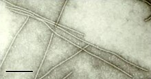
 Virionen von Carnation latent virus (CLV) | ||||||||||||||||||
| Systematik | ||||||||||||||||||
| ||||||||||||||||||
| Taxonomische Merkmale | ||||||||||||||||||
| ||||||||||||||||||
| Wissenschaftlicher Name | ||||||||||||||||||
| Carlavirus | ||||||||||||||||||
| Links | ||||||||||||||||||
|

Carlavirus, früher bekannt als Carnation latent virus group und Carlavirus group, ist eine Gattung von Viren in der Ordnung Tymovirales, Familie Betaflexiviridae, Unterfamilie Quinvirinae. Das Genom ist eine Einzelstrang-RNA mit positiver Polarität, (+)ssRNA (Gruppe IV der Baltimore-Klassifikation). Die Virionen (Viruspartikel) sind gerade oder leicht gewundene Filamente. Als natürliche Wirte dienen Pflanzen (Pflanzen- oder Phytoviren). Derzeit (Stand März 2021) gibt in dieser Gattung es vom International Committee on Taxonomy of Viruses (ICTV) bestätigt 53 Spezies (Arten), einschließlich der Typusart Carnation latent virus. Zu den mit dieser Gattung assoziierten Krankheiten gehören: Mosaik- und Ringspot-Symptome.[3][1]
Etymologie
Der Gattungsname Carlavirus ist eine Kombination aus der ersten Silbe der ersten beiden Wortteile im Namen der Typusart Carnation latent virus.
Beschreibung
Die Gattung Carlavirus wird im 9. Bericht des ICTV (2009) beschrieben.
Morphologie
Die Virionen (Virusteilchen) der Gattung sind nicht umhüllt, fadenförmig (filamentös) gerade oder leicht gebogen mit Helixsymmetrie. Ihre Länge beträgt grob 470–700 nm (im Extremfall 310–1000 nm, die Angaben schwanken je nach Autor) und 12–15 nm im Durchmesser bei einer Ganghöhe von etwa 3,4 nm.[4][3]
Genom

Das Genom besteht aus einem einzelsträngigen RNA-Molekül positiver Polarität von üblicherweise 7,4–7,7 kb (Kilobasen), ggf. auch (je nach Autor) 5,8–9,0 kb.[4][3]
Die Gattung zeichnet sich dadurch aus, dass sie sechs Offenen Leserahmen (Open Reading Frames, ORFs) mit kurzen untranslatierten Regionen (UTRs) an den Enden: Der 3'-Terminus ist polyadenyliert, bei einigen Arten ist das 5'-Ende mit einer Cap-Struktur versehen.[4][3]
Das Genom kodiert für 3 bis 6 Proteine, darunter ein Kapsidprotein, das sich am 3'-Ende befindet, und eine RNA-abhängige RNA-Polymerase, die sich am 5'-Ende des Genoms befindet. Beim Kartoffel-M-Virus kodiert ORF1 für ein Polypeptid mit 223 kDa (Kilodalton), das die Replikase des Virus darstellt.[4][3]
Die sich überlappenden Gene/ORFs 2, 3 und 4 bilden einen Block (Triple Gene Block, TGB) und kodieren für Polypeptide von 25, 12 und 7 kDa. Diese Triple-Gen-Block-Proteine (Triple Gene Block Proteins, TGBp: TGBp1, TGBp2 und TGBp3) ermöglichen oder erleichtern als Movement-Proteine die Bewegung der Virionen von Zelle zu Zelle und über große Distanzen. Einen solchen TGB findet man bei allen Betaflexiviridae mit Ausnahme der Gattungen Capillovirus, Citrivirus, Trichovirus, und Vitivirus, die ein einfaches Movement-Protein (MP) haben.[4][3]
ORF5 kodiert für ein Kapsidprotein (CP) mit 34 kDa und überlappt mit ORF6, der für ein Cystein-reichen Protein mit 11–16 kDa kodiert.[4][3]
RBP ist ein RNA-bindendes Protein:[3] ORF 5 überlappt mit ORF 6, der für ein Cystein-reichen Polypeptids mit 11–16 kDa kodiert, dessen Funktion noch nicht ganz geklärt ist. Seine Fähigkeit, Nukleinsäure zu binden, lässt vermuten, dass es die Übertragung durch Blattläuse erleichtern, oder am Gen-Silencing des Wirts oder der viralen RNA-Replikation beteiligt sein könnte.[4]
Replikationszyklus
Die virale Replikation erfolgt im Zytoplasma der Wirtszelle und ist lysogen. Der Eintritt in die Wirtszelle erfolgt durch Eindringen in diese. Die Replikation folgt dem Modell der Replikation von Einzelstrang-RNA-Viren positiver Polarität. Die Transkription erfolgt nach dem Modell von Einzelstrang-RNA-Viren positiver Polarität.
Übertragung
Die Viren werden durch Insekten (Blattläuse) als Vektoren übertragen.[4][3] Die Infektion wird manchmal durch Blattläuse in einem semi-persistenten Modus verbreitet, d. h. der Vektor ist für einige Stunden infektiös.[5] Einige Arten werden durch die Tabakmottenschildlaus (Bemisia tabaci) in einem semi-persistenten Modus oder durch das Saatgut übertragen.[6] Die meisten Arten infizieren nur einige wenige Wirte und verursachen Infektionen mit wenigen oder keinen Symptomen, z. B. American hop latent virus (AHLV) und Lily symptomless virus (LSV). Andere, wie das Blueberry scorch virus (BlSV) und das Poplar mosaic virus (PMV, Pappel-Mosaik-Virus), verursachen aber schwere Krankheiten.[7]
Systematik
Die Gattung wurde erstmals im ersten Bericht der ICTV 1971 als Carnation latent virus group vorgeschlagen, wurde aber 1975 in Carlavirus group und 1995 (6. Bericht) in die Gattung Carlavirus umbenannt. Im Jahr 2005 (8. Bericht) wurde es in die Familie der Flexiviridae gestellt, nachdem es zuvor nicht zugeordnet war.[8] Die aktuelle Position im 9. Bericht (2009) als Gattung der Familie Betaflexiviridae ergibt sich aus der späteren Unterteilung der Flexiviridae.[1]
Mit Stand Mai/Juni 2024 ist die Systematik der Gattung wie folgt:[9][10]
Ordnung Tymovirales
Familie Betaflexiviridae
● Unterfamilie Quinvirinae
- Gattung Carlavirus
- Spezies Carlavirus alphacapsici mit Pepper virus A (PepVA)
- Spezies Carlavirus alphacliviae mit Clivia carlavirus A (ClCVA)
- Spezies Carlavirus alphaconiti mit Aconite virus A (AcVA)
- Spezies Carlavirus alphaligustri mit Ligustrum virus A (LiVA, Ligustervirus A)
- Spezies Carlavirus alpharosae mit Rose virus A (RoVA)
- Spezies Carlavirus americanense mit American hop latent virus (AHLV)
- Spezies Carlavirus atractylodis mit Atractylodes mottle virus (AtrMV)
- Spezies Carlavirus betachrysanthemi mit Chrysanthemum virus B (CVB)
- Spezies Carlavirus betagapanthi mit Agapanthus carlavirus B (AgCVB)
- Spezies Carlavirus betaphlocis mit Phlox virus B (PhVB, Phloxvirus B)
- Spezies Carlavirus betarosae mit Rose virus B (RVB)
- Spezies Carlavirus betulae mit Birch carlavirus (BiCV)
- Spezies Carlavirus cacti mit Cactus virus 2 (CV2)


- Spezies Carlavirus chisolani mit Potato virus H (PotVH, PVH, Kartoffelvirus H)
- Spezies Carlavirus chloroipomoeae mit Sweet potato chlorotic fleck virus (SPCFV)
- Spezies Carlavirus colei mit Coleus vein necrosis virus (CVNV)
- Spezies Carlavirus cornutum mit Sint-jan's onion latent virus alias Sint-Jan onion latent virus (SJOLV, SjoLV, SJOLV0)
- Spezies Carlavirus cucumis mit Cucumber vein-clearing virus (CvCV)
- Spezies Carlavirus deltasambuci mit Sambucus virus D alias Elderberry carlavirus D (SVD, Holundervirus D)
- Spezies Carlavirus duocacti mit Cactus carlavirus 2 (CCV-2)
- Spezies Carlavirus duopseudostellariae mit Pseudostellaria heterophylla carlavirus 2 (PhCV2)
- Spezies Carlavirus epsilonsambuci mit Sambucus virus E alias Elderberry carlavirus E (SVE, Holundervirus E)
- Spezies Carlavirus fragariae mit Strawberry pseudo mild yellow edge virus (SPMYEV)
- Spezies Carlavirus gammasambuci mit Sambucus virus C alias Elderberry carlavirus C (SVC, Holundervirus C)
- Spezies Carlavirus hellebori mit Helleborus mosaic virus (HMV, Nieswurz-Mosaikvirus)
- Spezies Carlavirus humuli mit Hop mosaic virus (HpMV, Hopfen-Mosaikvirus)
- Spezies Carlavirus hydrangeae mit Hydrangea chlorotic mottle virus (HCMV)
- Spezies Carlavirus ipomoeae mit Sweet potato C6 virus (SPC6V, SPC6V0, Süßkartoffelvirus C6)
- Spezies Carlavirus jasmini mit Jasmine virus C (JVC)
- Spezies Carlavirus latensaconiti mit Aconitum latent virus (AcLV)
- Spezies Carlavirus latensallii mit Garlic common latent virus (GarCLV)
- Spezies Carlavirus latensascalonici mit Shallot latent virus (SLV, SLV000)
- Spezies Carlavirus latensbrassicae mit Cole latent virus (CoLV)
- Spezies Carlavirus latenscapparis mit Caper latent virus (CapLV)
- Spezies Carlavirus latensdianthi mit Carnation latent virus (CLV)[11]
- Spezies Carlavirus latensdioscoreae mit Yam latent virus (YLV)
- Spezies Carlavirus latensgaillardiae mit Gaillardia latent virus (GLV)
- Spezies Carlavirus latenshippeastri mit Hippeastrum latent virus (HLV)
- Spezies Carlavirus latenshumuli mit Hop latent virus (HpLV, unterscheide: Hop latent viroid, HpLVd, Gattung Cocadviroid)
- Spezies Carlavirus latenskalanchoe mit Kalanchoë latent virus (KLV)
- Spezies Carlavirus latensnarcissi mit Narcissus common latent virus (NCLV)
- Spezies Carlavirus latensnerinis mit Nerine latent virus (NeLV)
- Spezies Carlavirus latensolani mit Potato latent virus (PotLV)
- Spezies Carlavirus latenspassiflorae mit Passiflora latent virus (PLV)
- Spezies Carlavirus latensverbenae mit Verbena latent virus (VeLV)
- Spezies Carlavirus lilii mit Lily symptomless virus (LSLV, LSV, Lily-symptomless-Virus, Symptomloses Lilienvirus)
- Spezies Carlavirus maculapapayae mit Papaya mottle-associated virus (PapMaV)
- Spezies Carlavirus melonis mit Melon yellowing-associated virus (MYaV)
- Spezies Carlavirus miphlocis mit Phlox virus M (PhVM, Phloxvirus M)
- Spezies Carlavirus mirabilis mit Mirabilis jalapa mottle virus (MjMV)


- Spezies Carlavirus misolani mit Potato virus M (PotVM, PVM, Kartoffelvirus M)
- Spezies Carlavirus mitipapayae mit Papaya mild mottle associated virus (PaMMaV)
- Spezies Carlavirus necroligustri mit Ligustrum necrotic ringspot virus (LNRSV)
- Spezies Carlavirus necroretis mit Helleborus net necrosis virus (HNNV)
- Spezies Carlavirus oleraceae mit Cole mild mosaic virus (CoMMV)
- Spezies Carlavirus petasitis mit Butterbur mosaic virus (BuMV, Pestwurz-Mosaikvirus)
- Spezies Carlavirus pisi mit Pea streak virus (PeSV)
- Spezies Carlavirus pisolani mit Potato virus P (PotVP, PVP, Kartoffelvirus P)
- Spezies Carlavirus populi mit Poplar mosaic virus (PopMV, Pappel-Mosaikvirus)
- Spezies Carlavirus rhochrysanthemi mit Chrysanthemum virus R (CVR)
- Spezies Carlavirus sigmadaphnis mit Daphne virus S (DVS, Seidelbastvirus S)
- Spezies Carlavirus sigmahelenii mit Helenium virus S (HVS, Sonnenbrautvirus S)
- Spezies Carlavirus sigmaphlocis mit Phlox virus S (PhVS, Phloxvirus S)
- Spezies Carlavirus sigmascalonici mit Shallot virus S ShVS
- Spezies Carlavirus sigmasolani mit Potato virus S (PotVS, PVS, Kartoffelvirus S)
- Spezies Carlavirus sigmavaccinii mit Blueberry virus S (BVS)
- Spezies Carlavirus trifolii mit Red clover vein mosaic virus (RCVMV, Rotklee-Adernmosaikvirus)
- Spezies Carlavirus tripseudostellariae mit Pseudostellaria heterophylla carlavirus 3 (PhCV3)
- Spezies Carlavirus unicacti mit Cactus carlavirus 1 (CCV-1)
- Spezies Carlavirus uniglycinis mit Soybean carlavirus 1 (SCV1)
- Spezies Carlavirus unipseudostellariae mit Pseudostellaria heterophylla carlavirus 1 (PhCV1)
- Spezies Carlavirus unisteviae mit Stevia carlavirus 1 (StCV1)
- Spezies Carlavirus vaccinii mit Blueberry scorch virus (BlScV, BlSV)

- Spezies Carlavirus vignae mit Cowpea mild mottle virus (CPMMV)
Literatur
- Giovanni P. Martelli, Pasquale Saldarelli: Carlavirus. In: Christian Tidona, Gholamreza Darai (Hrsg.): The Springer Index of Viruses. Springer Science & Business Media, New York 2012, ISBN 978-0-387-95919-1, S. 521–532, doi:10.1007/978-0-387-95919-1_75 (eingeschränkte Vorschau in der Google-Buchsuche – Leseprobe).
- Suzanne Astier, Josette Albouy, Yves Maury: Principles of Plant Virology: Genome, Pathogenicity, Virus Ecology. Science Publishers, Enfield 2007, ISBN 978-1-57808-503-3, S. 78.
- Carlavirus Isolation and RNA Extraction. In: Gary D. Foster, Sally C. Taylor (Hrsg.): Plant Virology Protocols: From Virus Isolation to Transgenic Resistance. Humana Press, New York 1998, ISBN 0-89603-385-6, S. 145, doi:10.1385/0-89603-385-6:145.
- David Pimentel: Encyclopedia of Pest Management. Marcel Dekker, New York 2002, ISBN 0-8247-0632-3, S. 407 (eingeschränkte Vorschau in der Google-Buchsuche – Leseprobe).
- Andrew M. Q. King: Genus Carlavirus. In: Virus Taxonomy: Ninth Report of the International Committee on Taxonomy of Viruses. Ninth report of the International Committee on Taxonomy of Viruses. Elsevier, London 2012, ISBN 978-0-12-384684-6, S. 924 (eingeschränkte Vorschau in der Google-Buchsuche).
Weblinks
- Taxon: Genus Carlavirus (virus) The Taxonomicon
- Viralzone: Carlavirus
- ICTV (Seite nicht mehr abrufbar. Suche in Webarchiven)
Einzelnachweise
- ↑ a b c d e ICTV: ICTV Master Species List 2019.v1, New MSL including all taxa updates since the 2018b release, March 2020 (MSL #35)
- ↑ ICTV Master Species List 2018b v1 MSL #34, Feb. 2019
- ↑ a b c d e f g h i Viral Zone. ExPASy, abgerufen am 21. April 2021.
- ↑ a b c d e f g h ICTV: ICTV 9th Report (2011) – Betaflexiviridae
- ↑ David Pimentel: Encyclopedia of Pest Management. Marcel Dekker, New York 2002, ISBN 0-8247-0632-3, S. 407 (eingeschränkte Vorschau in der Google-Buchsuche – Leseprobe).
- ↑ Suzanne Astier, Josette Albouy, Yves Maury: Principles of Plant Virology: Genome, Pathogenicity, Virus Ecology. Science Publishers, Enfield 2007, ISBN 978-1-57808-503-3, S. 78.
- ↑ Carlavirus Isolation and RNA Extraction. In: Gary D. Foster, Sally C. Taylor (Hrsg.): Plant Virology Protocols: From Virus Isolation to Transgenic Resistance. Humana Press, New York 1998, ISBN 0-89603-385-6, S. 145, doi:10.1385/0-89603-385-6:145.
- ↑ M. J. Adams, J. F. Antoniw, M. Bar-Joseph, A. A. Brunt, T. Candresse, G. D. Foster, G. P. Martelli, R. G. Milne, S. K. Zavriev, C. M. Fauquet: The new plant virus family Flexiviridae and assessment of molecular criteria for species demarcation. In: Archives of Virology. Band 149, Nr. 5, Mai 2004, ISSN 0304-8608, S. 1045–1060, doi:10.1007/s00705-004-0304-0, PMID 15098118.
- ↑ ICTV: Taxonomy Browser.
- ↑ ICTV: Virus Metadata Resource (VMR).
- ↑ ICTVdB: Carnation latent virus (via WebArchiv vom 11. September 2006)
Auf dieser Seite verwendete Medien
Autor/Urheber: (of code) -xfi-, Lizenz: CC BY-SA 3.0
The Wikispecies logo created by Zephram Stark based on a concept design by Jeremykemp.
Autor/Urheber: R. G. Milne., Lizenz: CC BY-SA 4.0
Filamentous particles of an isolate of the species Carnation latent virus. The bar represents 100 nm.
Left, normal leaf of Nicotiana debneyi. Right, vein clearing caused in N. debneyi by Potato virus S (PVS), genus Carlavirus
Autor/Urheber: Brito et al, Lizenz: CC BY-SA 3.0
Transmission electron micrograph showing Cowpea mild mottle virus particles in sap of symptomatic V. unguiculata subsp. sesquipedalis leaf. Bar = 100 nm
Autor/Urheber: M. J. Adams, T. Candresse, J. Hammond, J. F. Kreuze, G. P. Martelli, S. Namba, M. N. Pearson, K. H. Ryu, P. Saldarelli, N. Yoshikawa, Lizenz: CC BY-SA 4.0
Genome organization of potato virus M showing the relative positions of the ORFs and their expression products. Mtr, methyltransferase; P-Pro, papain-like protease; Hel, helicase; RdRp, RNA-dependent RNA polymerase; CP, capsid protein; NB, nucleic acid binding protein. The 25K, 12K and 7K proteins constitute the triple gene block.
Autor/Urheber: Yuan-Yuan Li, Ru-Nan Zhang, Hai-Ying Xiang, Hesham Abouelnasr, Da-Wei Li, Jia-Lin Yu, Jenifer Huang McBeath, Cheng-Gui Han, Lizenz: CC BY-SA 4.0
Transmission electron micrograph showing the negative-stained virion of purified Potato virus H (PVH) virions.
Autor/Urheber: Yu ZHANG, Yan-ling GAO, Wan-qin HE, Ya-qin WANG, Lizenz: CC BY-SA 4.0
An electron micrograph of purified potato virus M (PVM) virions. The PVM virions were negatively stained with 1% phosphotungstic acid, pH 7.5, prior to photograph. Bar=0.2 μm.
Autor/Urheber: David Gent, USDA Agricultural Research Service, Bugwood.org, Lizenz: CC BY-SA 3.0
Carnation Latent Virus (CLV, genus Carlavirus). Host: common hop (Humulus lupulus L.)
Autor/Urheber: David Gent, USDA Agricultural Research Service, Bugwood.org, Lizenz: CC BY-SA 3.0
Carnation Latent Virus (CLV, genus Carlavirus) Host: common hop (Humulus lupulus L.)
Autor/Urheber: Yuan-Yuan Li, Ru-Nan Zhang, Hai-Ying Xiang, Hesham Abouelnasr, Da-Wei Li, Jia-Lin Yu, Jenifer Huang McBeath, Cheng-Gui Han, Lizenz: CC BY-SA 4.0
Genomic organization of Potato virus H (PVH). (A) Schematic diagram of the PVH genome. The solid line represents the RNA genome and the boxes represent the ORFs. The putative protein products are indicated. (B) Genome cloning strategy and locations of the cDNA clones used for PVH sequencing: p1 represents the RT-PCR-generated sequence using the degenerate primer during the detection of Potato leafroll virus; h1, h2 and h3 represent RT-PCR-generated sequences using degenerate primers specific to carlaviruses and PVH specific primers based on the p1 sequence; rc is the 5-terminal clone generated by 5' rapid amplification of cDNA ends.
Autor/Urheber: ViralZone, SIB Swiss Institute of Bioinformatics, Philippe Le Mercier et al., Lizenz: CC BY 4.0
Genomkarte der Gattung Carlavirus


