CD66a
| Carcinoembryonic antigen-related cell adhesion molecule 1 | ||
|---|---|---|
 | ||
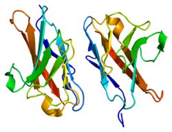
| nach PDB 2GK2 | ||
| Andere Namen | Biliary glycoprotein 1 | |
| Eigenschaften des menschlichen Proteins | ||
| Masse/Länge Primärstruktur | 526 Aminosäuren, 57.560 Da | |
| Bezeichner | ||
| Externe IDs | ||
| Vorkommen | ||
| Homologie-Familie | Hovergen | |
| Orthologe (Mensch) | ||
| Entrez | 634 | |
| Ensembl | ENSG00000079385 | |
| UniProt | P13688 | |
| Refseq (mRNA) | NM_001024912.2 | |
| Refseq (Protein) | NP_001020083.1 | |
| PubMed-Suche | 634 | |
Carcinoembryonic antigen-related cell adhesion molecule 1 (synonym CEACAM1, CD66a) ist ein Oberflächenprotein und Zelladhäsionsmolekül aus der Immunglobulin-Superfamilie.
Eigenschaften
CD66a ist eines von zwölf Vertretern der CEACAM.[1] Es wird in mehreren Isoformen in Endothelzellen, Epithelzellen, Granulozyten und Lymphozyten gebildet, von denen diejenigen mit langem zytosolischen Anteil am häufigsten vorkommen.[2] Isoform 1 ist an der Zelladhäsion, der Angiogenese und der Hemmung der Immunantwort beteiligt. Die Hemmung der Immunantwort erfolgt durch Zellkontakte mit anderen Zellen und eine Hemmung der Degranulation von zytotoxischen T-Zellen, eine Hemmung von NK-Zellen durch Bindung an andere CD66a und eine Hemmung der Bildung von Interleukin-1beta in Neutrophilen. Es ist glykosyliert und phosphoryliert.
CD66a bindet an PTPN11[3] und Annexin A2.[4]
CD66a ist an der Entstehung von Melanomen beteiligt.[5] Das Verhältnis der Isoformen CEACAM1-S/CEACAM1-L ist ein Prognosemarker bei einem nichtkleinzelligen Lungenkrebs.[6]
Weblinks
Einzelnachweise
- ↑ N. Beauchemin, A. Arabzadeh: Carcinoembryonic antigen-related cell adhesion molecules (CEACAMs) in cancer progression and metastasis. In: Cancer metastasis reviews. Band 32, Nummer 3–4, Dezember 2013, S. 643–671, doi:10.1007/s10555-013-9444-6, PMID 23903773.
- ↑ Z. Chen, L. Chen, S. W. Qiao, T. Nagaishi, R. S. Blumberg: Carcinoembryonic antigen-related cell adhesion molecule 1 inhibits proximal TCR signaling by targeting ZAP-70. In: Journal of Immunology. Band 180, Nummer 9, Mai 2008, S. 6085–6093, PMID 18424730.
- ↑ Huber M, Izzi L, Grondin P, Houde C, Kunath T, Veillette A, Beauchemin N: The carboxyl-terminal region of biliary glycoprotein controls its tyrosine phosphorylation and association with protein-tyrosine phosphatases SHP-1 and SHP-2 in epithelial cells. In: The Journal of Biological Chemistry. 274. Jahrgang, Nr. 1, Januar 1999, S. 335–44, doi:10.1074/jbc.274.1.335, PMID 9867848.
- ↑ Kirshner J, Schumann D, Shively JE: CEACAM1, a cell-cell adhesion molecule, directly associates with annexin II in a three-dimensional model of mammary morphogenesis. In: The Journal of Biological Chemistry. 278. Jahrgang, Nr. 50, Dezember 2003, S. 50338–45, doi:10.1074/jbc.M309115200, PMID 14522961.
- ↑ G. Turcu, R. I. Nedelcu, D. A. Ion, A. Brînzea, M. D. Cioplea, L. B. Jilaveanu, S. A. Zurac: CEACAM1: Expression and Role in Melanocyte Transformation. In: Disease markers. Band 2016, 2016, S. 9406319, doi:10.1155/2016/9406319, PMID 27642217, PMC 5013198 (freier Volltext).
- ↑ Y. Ling, J. Wang, L. Wang, J. Hou, P. Qian, W. Xiang-dong: Roles of CEACAM1 in cell communication and signaling of lung cancer and other diseases. In: Cancer metastasis reviews. Band 34, Nummer 2, Juni 2015, S. 347–357, doi:10.1007/s10555-015-9569-x, PMID 26081722.
Auf dieser Seite verwendete Medien
Autor/Urheber: Emw, Lizenz: CC BY-SA 3.0
Structure of the CEACAM1 protein. Based on PyMOL rendering of PDB 2gk2.
