Actinophrys sol
| Actinophrys sol | ||||||||||||
|---|---|---|---|---|---|---|---|---|---|---|---|---|
.jpg) Phasenkontrast-Bild von A. sol, | ||||||||||||
| Systematik | ||||||||||||
| ||||||||||||
| Wissenschaftlicher Name der Gattung | ||||||||||||
| Actinophrys | ||||||||||||
| Ehrenberg, 1830 | ||||||||||||
| Wissenschaftlicher Name der Art | ||||||||||||
| Actinophrys sol | ||||||||||||
| (O.F.Müller) Ehrenberg[1] |
-1.jpg)

Actinophrys sol ist eine Spezies (Art) eukaryotischer Einzeller (Protisten), die man früher als Heliozoa (Sonnentierchen, englisch sun animalcules) klassifizierte.[2] Da die Heliozoa heute allgemein nicht mehr als monophyletisch gelten, existiert diese Bezeichnung nur noch als informeller Name (morphologisch orientierter Sammelbegriff, kein Taxon). In der modernen Taxonomie gilt die Familie Actinophryidae (Gattung Actinophrys und die nah verwandte Gattung Actinosphaerium mit der Spezies Actinosphaerium eichhornii[3]) meistens in die Klade der Actinophryida, die von einigen Taxonomen im Rang einer Ordnung gesehen wird. Diese Actinophryida werden als Mitglieder der Raphidophyceae zu den Stramenopilen (alias Chromista oder Heterokonta) gestellt.[1] Andere Taxonomien gruppieren die Familie Actinophryidae letztlich ebenfalls zu den Chromista, wenn auch über andere Zwischenstufen[4] oder lassen diese gleich ganz offen.[5]
Die Actinophrys-Zellen sind annähernd kugelförmige (sphärisch-symmetrische) Einzeller mit einem großen zentralen Zellkern. Sie können nach der Nahrungsaufnahme eine äußere (periphere) Schicht von Nahrungsvakuolen aufweisen. Sie besitzen Mikrotubuli-Arrays (Axoneme), die ⁹innen am Zellkern enden.[6]
Der Lebensbereich von Actinophrys ist das Süßwasser.[6] Sie schwimmen frei in stehenden oder langsam fließenden Gewässern.[3]
Actinophrys sol ist Einzeller des Jahres 2013.[7][8]
Beschreibung
Die Einzeller der Gattung Actinophrys sind räuberische Organismen. Man findet sie vor allem im Süßwasser, wo sie im offenen Wasser zwischen Schilf und fadenförmigen Algen schwimmen.[9] Die Actinophrys-Zellen zeichnen sich dadurch aus, dass sie im Gegensatz zu einigen verwandten Gattungen nur einen einzigen Zellkern haben.[10] Wie die anderen informell als Sonnentierchen bezeichneten Einzeller sind sie (annähernd) kugelförmig und sind häufig von einer Schale aus Silikat oder organischem Material umhüllt. Die Einzeller scheiden diese Schale in Form von Schuppen oder Platten in einer gallertartigen Hülle aus. Die Schalen weisen in der Familie Actinophryidae eine große Vielfalt an Formen auf, die zur Artbestimmung herangezogen werden können. Die Actinophrys-Zellen pflanzen sich ungeschlechtlich durch binäre Spaltung (Schizotomie), evtl. auch durch Knospung fort.[9]
Die Actinophrys-Zellen besitzen spezialisierte Pseudopodien (Scheinfüßchen), die Axopodien genannt werden, da sie sich strahlenförmig nach außen erstrecken. Diese Anhängsel dienen der Fortbewegung und dem Einfangen von Nahrung (Protozoen, Mikroalgen und andere kleine Organismen). Die Axopodien bestehen aus einem zentralen Kern, der ein Bündel von Mikrotubuli enthält und von einer äußeren Schicht aus fließendem Zytoplasma umgeben ist. Die Mikrotubuli sind in spezifischen Mustern vernetzt, die von Art zu Art variieren. Das äußere Zytoplasma (Ektoplasma) kann spezielle Organellen enthalten, die zum Fangen von Beutetieren herausschnellen können.[9]
Die gleichberechtigte Konjugation wurde von F. Schaudinn bei Actinophrys vollständig demonstriert: zwei Individuen nähern sich an, treten in engen Kontakt und sind von einer gemeinsamen Zystenwand umgeben. Der Kern eines der beiden ‚Männchen‘ teilt sich, und ein Kern geht auf beiden Seiten an die Oberfläche und wird mit einem kleinen Teil des Zytoplasmas als abortive (dem Tod geweihte) Zelle abgespalten; die beiden verbleibenden Kerne, die in der zellulären Beziehung „Cousins ersten Grades“ sind, verschmelzen nun, wie es auch bei den Zytoplasten (Zellmembran und Zytoplasma) der Fall ist. Die so entstandene gekoppelte Zelle oder Zygote teilt sich in zwei Teile, die sich wiederum verklumpen.[3]
Systematik
Quellen der folgenden Systematik sind:
- (A) — AlgaeBase[1]
- (I) — Integrated Taxonomic Information System (ITIS)
- (M) — Microworld[6]
- (N) — National Center for Biotechnology Information (NCBI) Taxonomy Browser[5]
- (W) — World Register of Marine Species (WoRMS)[4]
- (µ) — MicroscopyU, Nikon[9]
Nachgestellt ‚:U‘ deutet eine gemäß der jeweiligen Quelle nicht voll gültige Charakterisierung an.
Familie: Actinophryidae Dujardin 1841 (A,I,M,N)
- Gattung: Actinophrys Ehrenberg 1830 (A,I,M,N,W,µ)
- Spezies: Actinophrys pontica Valkanov 1940 (A,M)
- Spezies: Actinophrys salsuginosa D.&nbso;J. Patterson 2001 (A,M)
- Spezies: Actinophrys sol (O. F. Müller, 1773) Ehrenberg 1830/1840 (A:U,I,M,N,W:U) — Typusart (M)
- A. sol green Angie Opitz 2015 (M) — in einer Probe aus einem Torfmoor in Tirol. (M)
- Spezies: Actinophrys tauryanini (Mikrjukov, 1996) Mikrjukov & D.J.Patterson 2001 (A:U)
- Spezies: Actinophrys vesiculata Pen. (I)
Verschiebungen (Auswahl): Actinophrys viridis ist jetzt Acanthocystis turfacea (siehe Acanthocystis)
Ein Teil der früher Actinophrys sol zugerechneten Beobachtungen bzw. Varianten werden nach Curds (1986) als Podophrya fixa (O. F. Müller, 1786) Ehrenberg, 1833 klassifiziert.[11][12][13] Diese Spezies ist zu unterscheiden von Podophrya sol (Mechnikov, 1864) n. comb. [früher auch bezeichnet als Sphaerophrya sol Mechnikov, 1864].[11] Da die einzelnen Spezies jeweils nur von einem Teil der Quellen bestätigt werden, muss die Artenliste insgesamt als unsicher gelten.
Etymologie
Der Artname Actinophrys setzt sich zusammen aus neulateinisch actin von altgriechisch ἀκτίςaktis, deutsch ‚Strahl‘[14] und ὀφρύςophrýs, deutsch ‚Augenbraue‘,[15][16] ‚Rand‘, bedeutet also frei übersetzt ‚Strahlenkranz‘.
Bildergalerie
 Actinophrys sol, Illustration (1852)
 A. sol bei der Nahrungsaufnahme. Illustration (1852)
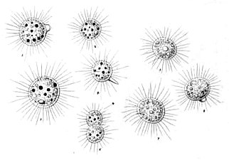
 A. sol aus Brehms Tierleben (1918)
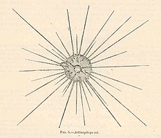

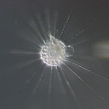 Mikrophotographie von A. sol aus einer Probe vom Ryugaoka-Park, Ryūgasaki, Präfektur Ibaraki, Japan
 Die Mikrophotographie von A. sol zeigt die annähernd sphärische Symmetrie.
 A. sol, Zellverband mit zwei gemeinsamen Nahrungsvakuolen, DIK-Aufnahme.
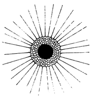
 A. sol, Illustration von 1830
Weblinks
- Flyer Actinophrys sol – Einzeller des Jahres 2013 (www.protozoologie.de).
- Actinophrys sol: Einzeller des Jahres 2013. In: Mikrokosmos, Heft 1, 102. Jahrgang, Januar 2013, S. 15, ISSN 0026-3680.
- LifeGate: Centrohelea – Actinophrys.
Einzelnachweise
- ↑ a b c d AlgaeBase: Actinophrys Ehrenberg, 1830 (Genus).
- ↑ ITIS: Actinophrys, Integrated Taxonomic Information System.
- ↑ a b c Heliozoa. In: Encyclopædia Britannica, 1911 (WikiSource).
- ↑ a b WoRMS: Actinophrys (Genus).
- ↑ a b NCBI Taxonomy Browser: Actinophrys, Detail: Actinophrys (genus), graphisch: Actinophrys, Lifemap NCBI Version.
- ↑ a b c Microworld: Genus Actinophrys Ehrenberg, 1830 und Actinophryidae, World of amoeboid organisms (arcella.nl). Mit Bildern, insbes. von A. sol.
- ↑ Flyer Actinophrys sol ( des vom 28. August 2016 im Internet Archive) Info: Der Archivlink wurde automatisch eingesetzt und noch nicht geprüft. Bitte prüfe Original- und Archivlink gemäß Anleitung und entferne dann diesen Hinweis. – Einzeller des Jahres 2013 (www.protozoologie.de).
- ↑ Actinophrys sol: Einzeller des Jahres 2013. In: Mikrokosmos, Heft 1, 102. Jahrgang, Januar 2013, S. 15, ISSN 0026-3680.
- ↑ a b c d Actinophrys (Protozoan) Videos, Differential Interference Contrast (DIC) Pond Life Video Gallery, Nikon.
- ↑ Eiji Kinoshita, Yoshinobu Shigenaka, Toshinobu Suzaki: The ultrastructure of contractile tubules in the heliozoon Actinophrys sol and their possible involvement in rapid axopodial contraction. In: The Journal of Eukaryotic Microbiology. 48. Jahrgang, Nr. 5, 2001, S. 519–526, doi:10.1111/j.1550-7408.2001.tb00187.x, PMID 11596916 (englisch, wiley.com).
- ↑ a b Colin Robert Curds: A revision of the Suctoria (Ciliophora, Kinetofragminophora) 4. Podophrya and its morphological relatives. In: Bulletin of the British Museum (Natural History) Zoology, Band 50, London, BM(NH), 1986, S. 59–91; BioStor:38, PDF (archive.org), BHL:8103 (englisch). Siehe insbes. S. 62 und Fig. 28.
- ↑ NCBI Taxonomy Browser: Podophrya fixa (species).
- ↑ WoRMS: Podophrya fixa O. F. Muller, 1786 (Species).
- ↑ Renate Wahrig-Burfeind (Hrsg.): Wahrig. Illustriertes Wörterbuch der deutschen Sprache. ADAC-Verlag, München 2004, ISBN 3-577-10051-6, S. 39.
- ↑ Ludwig August Kraus: Kritisch-etymologisches medicinisches Lexikon, 3. Auflage, Verlag der Deuerlich- und Dieterichschen Buchhandlung, Göttingen 1844, S. 692 und 1072.
- ↑ actinophrys noun, Merriam-Webster.
Auf dieser Seite verwendete Medien

Autor/Urheber: ja:User:NEON / User:NEON_ja, Lizenz: CC BY-SA 3.0
Actinophrys sol / from Ryugaoka park, Ryūgasaki, Ibaraki Pref., Japan / Microscope:Leica DMRD (DIC)
_(14576779497).jpg)
Autor/Urheber: Internet Archive Book Images, Lizenz: No restrictions
Identifier: inbrookbayouorli00bayl (find matches)
Title: In brook and bayou; or, Life in the still waters
Year: 1897 (1890s)
Authors: Bayliss, Clara Kern, 1848-
Subjects: Zoology
Publisher: New York, D. Appleton and company
Contributing Library: The Library of Congress
Digitizing Sponsor: The Library of Congress
View Book Page: Book Viewer
About This Book: Catalog Entry
View All Images: All Images From Book
Click here to view book online to see this illustration in context in a browseable online version of this book.
Text Appearing Before Image:
his hat and has to crawl out andlie around unclothed till he can make another(Chart I, Fig. 5) ! III. THE SUN ANIMALCULE. (Actinophrys sol.) If you blow soap bubbles from the end ofa tube into the air, blowing carefully with fre-quent pauses, you may make, not a single bub-ble, but a ball of small bubbles. Now, if you can imagine that out from thisglobe of bubbles, radiating in all directions, arespines as colorless as the bubbles themselves,and that every moment or two a large bubblebursts and then forms itself again, you willknow how the Actinophrys sol looks. It is called the sun animalcule because therays from the ball make it look like the oldpictures of the sun. You can find it in jouv jardiniere and every-where in fresh water, where other microscopicanimals live, but its favorite residence is onsphagnum or bog moss. 20 IN BROOK AND BAYOU. A large one is hr of an incli in diameter. 65 0 It is very quiet and well behaved. It has agently gliding or floating motion, and moves in
Text Appearing After Image:
Fig. 6.—1, Pulsating vacuole; 2, food in food-vacuole. a circular course when it moves at all. But itremains in one place for long periods of time. It can travel, but seldom does. It can eat,but seldom does. It can withdraw its rays,and flatten itself like an amoeba, but seldomdoes. Although it remains so quiet that one hasfull opportunity for observing it, little is knownof its life history. It is beautiful and nothingmore ; and so it lives, and nothing more, re- RHIZOPODS. 21 minding one of the question tlie page put toBrutuss wife : What shaU I do ? Run to the Capitol, and nothingmore ?And so return, and nothing more ? But beauty to be really interesting must becoupled witli energy and vivacity, and the sunanimalcule has little of either. Yet it is fond of society, and is often seenclosely associated with others, their spines in-terlaced, the animacules piled in a heap, some-times to the number of fifteen in one colony. The actinophrys multiplies by division, andthe colonies are
Note About Images

Autor/Urheber: Michael Linnenbach, Lizenz: CC BY-SA 3.0
Heliozoen (Actinophrys sol) - Zellverband mit zwei gemeinsamen Nahrungsvakuolen. (Lichtmikroskopie, DIK - Differentialinterferenzkontrast)
.jpg)
Autor/Urheber: User:Esseh~commonswiki. Original provided by User:Jeffdelonge as Actinophrys_sol.jpg, Lizenz: CC BY-SA 3.0
Microphotography showing pherical symmetry of Actinophrys sol

Illustration von Actinophrs sol. Detaillierter Text unter https://www.biodiversitylibrary.org/page/29684577#page/114/mode/1up (IVte Gruppe)
Autor/Urheber: Djpmapfer, Lizenz: CC BY-SA 4.0
Actinophrys is a uninculeate actinophryid heliozoon. It has a central nucleus. Microtubular arrays, axonemes, that support the arms of the cell terminate on the nucleus. Phase contrast microscopy.


Sonnentierchen (Actinophrys sol)
Autor/Urheber: Djpmapleferryman, Lizenz: CC BY-SA 4.0
Actinoiphrys sol, scanning electron micrograph of resting cyst showing irregular siliceous plates.

Sonnentierchen (Actinophrys sol) bei der Verdauung
Actinophrys sol Ehrenberg. a: in einer großen Nahrungsvakuole liegendes Nahrungspartikel; b: inneres feinkörniges Protoplasma (Endoplasma); c: axiales Filament eines Pseudopodiums, das sich nach innen zum Kern erstreckt; d: der zentrale Kern; e: kontraktile Vakuole; f: äußeres stark vakuolisiertes Protoplasma (Ektoplasma).
_(20226446908)_3.jpg)
Autor/Urheber: Brehm, Alfred Edmund, 1829-1884 (cropped & digitaly contrast enhanced grayscale), Lizenz: No restrictions
Einzeller : Actinophrys sol Ehrbg.

Autor/Urheber: M. Linnenbach, Lizenz: CC BY-SA 3.0
In der Nahrungsvakuole eines Sonnentierchens (Heliozoa) der Spezies Actinophrys sol eingeschlossenes Pantoffeltierchen (Paramecium) - Lichtmikroskopie, Differential-Interferenz-Kontrast (DIK)