Human brain anterior-inferior view description
John A Beal, PhD
Dep't. of Cellular Biology & Anatomy, Louisiana State University Health Sciences Center Shreveport
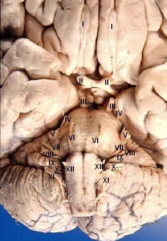
Human brain - anterior-inferior view - cerebral nerves
I. N. olfactorius - The fila olfactoria (approximately 20 nerve rootlets on each side) comprise the Olfactory nerves (I) which run from the olfactory receptors in the nasal cavity to the olfactory bulb.
II. N. opticus - The Optic nerves (II) are large & join at the midline to form the optic chiasm, then continue laterally as the optic tracts. The optic nerve is not a true nerve but rather a CNS tract.
III. N. oculomotorius - The Oculomotor nerves (III) emerge from a depression in the midbrain, the interpeduncular fossa, just caudal to the optic chiasm.
IV. N. trochlearis - The Trochlear nerves (IV) are small & are seen on the lateral surface of the midbrain. They are the only nerves which exit from the posterior side of the brain.
V. N. trigeminus - The Trigeminal nerves (V) are large and emanate from the lateral surface of the pons.
VI. N. abducens - The Abducens nerves (VI) exit near the midline from the inferior pontine sulcus which separates the pons from the medulla.
VII. N. facialis - Moving laterally in the inferior pontine sulcus, the Facial nerves (VII) can be seen.
VIII. N. vestibulocochlearis - Slightly lateral to the facial nerve is the Vestibulocochlear nerve (VIII).
IX. N. glossopharyngeus - The postolivary sulcus is a groove running rostrocaudally on the lateral surface of the medulla. From this sulcus pass the small Glossopharyngeal nerves (IX) rostrally and...
X. N. vagus - ...the much larger Vagus nerves (X) caudally.
XI. N. accessorius - The Spinal Accessory nerves (XI) exit the cervical cord then pass rostrally through the foramen magnum to exit the cranial vault with the lossopharyngeal and vagus nerves.
XII. N. hypoglossus - The Hypoglossal nerves (XII) exit the medulla via the preolivary sulcus.


Relevante Bilder


Relevante Artikel
HirnnervAls Hirnnerven werden jene Nerven bezeichnet, die direkt aus spezialisierten Nervenzellansammlungen im Gehirn entspringen, den Hirnnervenkernen, zumeist im Hirnstamm. .. weiterlesen